. 2008 Jun;17(6):1025-34.
doi: 10.1110/ps.073326608. Epub 2008 Apr 23.

The PIP2 binding mode of the C2 domains of rabphilin-3A

Affiliations

The PIP2 binding mode of the C2 domains of rabphilin-3A

Pierre Montaville et al. Protein Sci. 2008 Jun.

Abstract

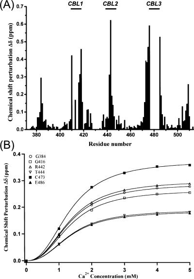
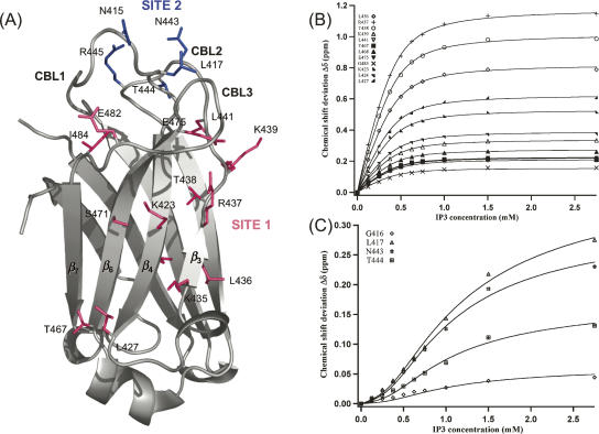
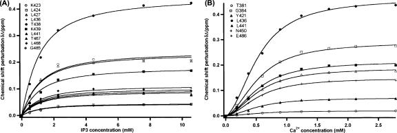
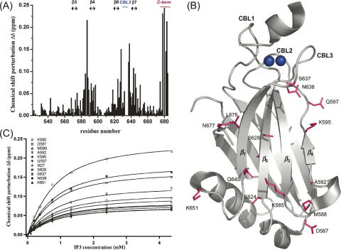
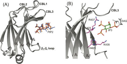
Phosphatidylinositol-4,5-bisphosphate (PIP2) is a key player in the neurotransmitter release process. Rabphilin-3A is a neuronal C2 domain tandem containing protein that is involved in this process. Both its C2 domains (C2A and C2B) are able to bind PIP2. The investigation of the interactions of the two C2 domains with the PIP2 headgroup IP3 (inositol-1,4,5-trisphosphate) by NMR showed that a well-defined binding site can be described on the concave surface of each domain. The binding modes of the two domains are different. The binding of IP3 to the C2A domain is strongly enhanced by Ca(2+) and is characterized by a K(D) of 55 microM in the presence of a saturating concentration of Ca(2+) (5 mM). Reciprocally, the binding of IP3 increases the apparent Ca(2+)-binding affinity of the C2A domain in agreement with a Target-Activated Messenger Affinity (TAMA) mechanism. The C2B domain binds IP3 in a Ca(2+)-independent fashion with low affinity. These different PIP2 headgroup recognition modes suggest that PIP2 is a target of the C2A domain of rabphilin-3A while this phospholipid is an effector of the C2B domain.


Figures

Figure 1.
Figure 1.
Structural organization of rabphilin-3A. The bar graph depicts the domains of rabphilin-3A whose three-dimensional structures have been solved: the Rab binding domain, the C2A domain, and the C2B domain.
Figure 2.
Figure 2.
Ca2+-binding properties of the C2A domain of rabphilin-3A. (A) 1H-15N chemical shift perturbations δΔ (see Materials and Methods) plotted against the sequence of the C2A domain fragment (371–510) upon addition of 5 mM Ca2+. Missing chemical shift deviations are associated with cross-peaks in intermediate chemical exchange rates at pH 7.0 in the absence of Ca2+ and with proline residues. The positions of the three Ca2+-binding loops (CBL1, CBL2, and CBL3) are highlighted on top. (B) Simultaneous fit performed on the chemical shift perturbations of six residue backbone HN cross-peaks according to the Ca2+ concentration using the Hill equation (Equation 3; see Materials and Methods).
Figure 3.
Figure 3.
IP3-binding mode of the C2A domain of rabphilin-3A. (A) Residues whose 1H-15N cross-peaks experienced significant chemical shift perturbations in the presence of 2.75 mM IP3 and 5 mM Ca2+ are mapped on the C2A domain crystal structure (PDB access number 2chd). Residues defining site 1 are colored in magenta; residues defining site 2 are colored in blue. (B) Simultaneous fit performed on chemical shift perturbation of 12 residue backbone HN cross-peaks from IP3-binding site 1 upon IP3 titration in the presence of 5 mM Ca2+ using Equation 1 (see Materials and Methods). (C) Simultaneous fit performed on chemical shift perturbation of four residue backbone HN cross-peaks upon IP3 titration in the presence of 5 mM Ca2+ using Equation 2 (see Materials and Methods). These residues correspond to the second IP3-binding site (site 2).
Figure 4.
Figure 4.
Ca2+ dependency of IP3-binding to the C2A domain. (A) Simultaneous fit performed on chemical shift perturbation of 10 residue backbone HN cross-peaks from IP3-binding site 1 upon IP3 titration without addition of Ca2+ to the buffer using Equation 1 (see Materials and Methods). (B) Simultaneous fit performed on the chemical shift perturbations of seven residue backbone HN cross-peaks according to the Ca2+ concentration using the Hill equation (Equation 3; see Materials and Methods) in the presence of 10 mM IP3.
Figure 5.
Figure 5.
IP3-binding mode of the C2B domain of rabphilin-3A. (A) 1H-15N chemical shift perturbations δΔ (see Materials and Methods) plotted against the sequence of the C2B domain fragment (519–684) upon the addition of 4.3 mM IP3 in the presence of 1 mM Ca2+. The secondary structure elements involved in the binding, β-strands 3, 4, 6, and 7, the Ca2+-binding loop 3, and the C-terminal tail are highlighted on top. (B) Residues experiencing 1H-15N chemical shift perturbations larger than 0.07 ppm upon IP3 titration are mapped on the C2B crystal structure (PDB accession number 2cm6, chain B). (C) Simultaneous fit performed on chemical shift perturbations of 11 residue backbone HN cross-peaks according to IP3 concentration using Equation 1 (see Materials and Methods).
Figure 6.
Figure 6.
Ca2+-independent IP3-binding to the C2B domain. Overlay of a region of 1H-15N HSQC C2B domain spectra. Red resonances correspond to the Ca2+-bound C2B domain (1 mM Ca2+), blue resonances to the Ca2+ free C2B domain (10 mM EGTA), magenta resonances to the Ca2+- and IP3-bound C2B domain (1 mM Ca2+, 4 mM IP3), and cyan resonances to the Ca2+ free, IP3-bound C2B domain (10 mM EGTA, 4 mM IP3). The black arrows indicate the chemical shift deviations of the cross-peaks (Ca2+ free and bound) upon IP3-binding.
Figure 7.
Figure 7.
Docking model for the C2A/PIP2 complex. (A) Ribbon representation of the final ensemble of models for the C2A/PIP2 complex. PIP2 is shown in colored stick representation. (B) Ribbon representation of the most representative model of the C2A/PIP2 complex. Side chains of residues actively involved in the binding model are represented in sticks and colored in magenta and blue; the IP3 carbon ring of the PIP2 molecule is colored in green; and the DAG chain is colored in light gray.

References

    1. Aikawa, Y., Xia, X., Martin, T.F. SNAP25, but not syntaxin 1A, recycles via an ARF6-regulated pathway in neuroendocrine cells. Mol. Biol. Cell. 2006;17:711–722. - PMC - PubMed
    1. Auguin, D., Barthe, P., Royer, C., Stern, M.H., Noguchi, M., Arold, S.T., Roumestand, C. Structural basis for the co-activation of protein kinase B by T-cell leukemia-1 (TCL1) family proto-oncoproteins. J. Biol. Chem. 2004;279:35890–35902. - PubMed
    1. Bai, J., Tucker, W.C., Chapman, E.R. PIP2 increases the speed of response of synaptotagmin and steers its membrane-penetration activity toward the plasma membrane. Nat. Struct. Mol. Biol. 2004;11:36–44. - PubMed
    1. Baldini, G., Martelli, A.M., Tabellini, G., Horn, C., Machaca, K., Narducci, P., Baldini, G. Rabphilin localizes with the cell actin cytoskeleton and stimulates association of granules with F-actin cross-linked by α-actinin. J. Biol. Chem. 2005;280:34974–34984. - PubMed
    1. Biadene, M., Montaville, P., Sheldrick, G.M., Becker, S. Structure of the C2A domain of rabphilin-3A. Acta Crystallogr. D Biol. Crystallogr. 2006;62:793–799. - PubMed

Publication types

MeSH terms

LinkOut - more resources