

Sex-specific differences in expression of histone demethylases Utx and Uty in mouse brain and neurons
- PMID: 18434530
- PMCID: PMC2643472
- DOI: 10.1523/JNEUROSCI.5382-07.2008
Sex-specific differences in expression of histone demethylases Utx and Uty in mouse brain and neurons
Abstract
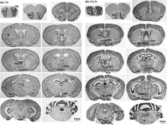
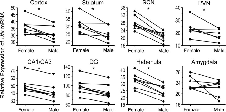
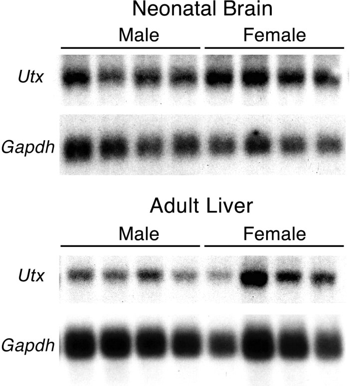
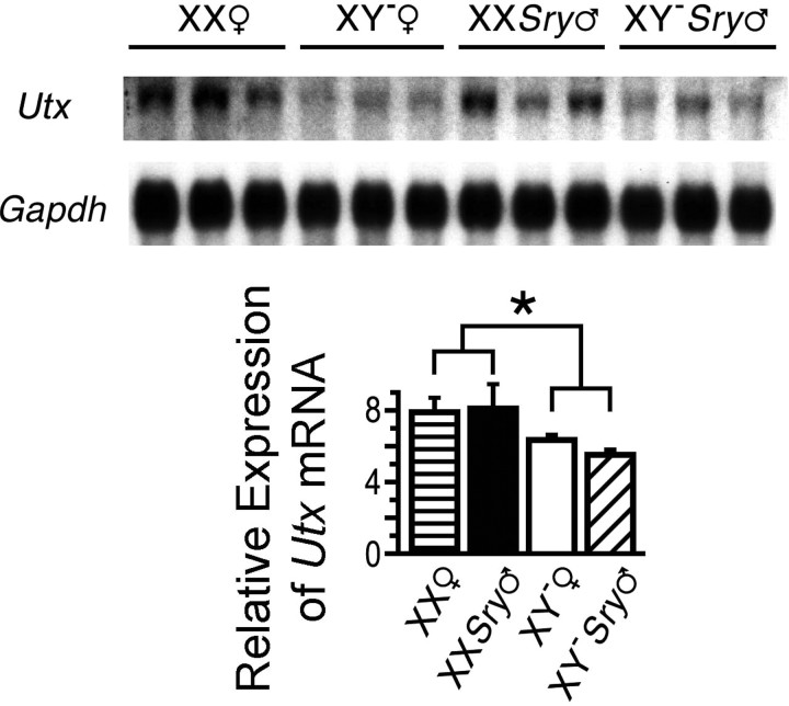
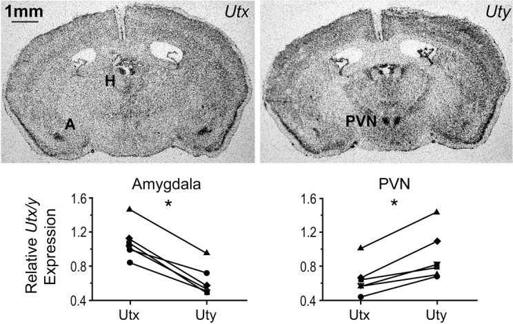
Although X inactivation is thought to balance gene expression between the sexes, some genes escape inactivation, potentially contributing to differences between males and females. Utx (ubiquitously transcribed tetratricopeptide repeat gene on X chromosome) is an escapee gene that encodes a demethylase specific for lysine 27 of histone H3, a mark of repressed chromatin. We found Utx to be expressed higher in females than in males in developing and adult brains and in adult liver. XX mice had a higher level of Utx than XY mice, regardless of whether they had testes or ovaries, indicating that the sexually dimorphic gene expression was a consequence of the sex chromosome complement. Females had significantly higher levels of Utx than males in most brain regions except in the amygdala. The regional expression of the Y-linked paralogue Uty (ubiquitously transcribed tetratricopeptide repeat gene on Y chromosome) was somewhat distinct from that of Utx, specifically in the paraventricular nucleus of the hypothalamus (high Uty) and the amygdala (high Utx), implying that the two paralogues may be differentially regulated. Higher expression of Utx compared with Uty was detected in P19 pluripotent embryonic carcinoma cells as well as in P19-derived neurons. This transcriptional divergence between the two paralogues was associated with high levels of histone H3 lysine 4 dimethylation at the Utx promoter and of histone H4 lysine 16 acetylation throughout the gene body, which suggests that epigenetic mechanisms control differential expression of paralogous genes.
Figures
References
-
- Adolphs R. Cognitive neuroscience of human social behaviour. Nat Rev Neurosci. 2003;3:165–178. - PubMed
-
- Agger K, Cloos PA, Christensen J, Pasini D, Rose S, Rappsilber J, Issaeva I, Canaani E, Salcini AE, Helin K. UTX and JMJD3 are histone H3K27 demethylases involved in HOX gene regulation and development. Nature. 2007;449:731–734. - PubMed
-
- Anderson AK, Phelps EA. Expression without recognition: contributions of the human amygdala to emotional communication. Psychol Sci. 2000;11:106–111. - PubMed
-
- Arnold AP. Sex chromosomes and brain gender. Nat Rev Neurosci. 2004;5:701–708. - PubMed
-
- Arnold AP, Gorski RA. Gonadal steroid induction of structural sex differences in the central nervous system. Annu Rev Neurosci. 1984;7:413–442. - PubMed
Publication types
MeSH terms
Substances
Grants and funding
LinkOut - more resources
Full Text Sources