Comparative Study
. 2008 Apr 23;28(17):4551-60.
doi: 10.1523/JNEUROSCI.5694-07.2008.

Isoflurane-induced caspase-3 activation is dependent on cytosolic calcium and can be attenuated by memantine

Affiliations
Comparative Study

Isoflurane-induced caspase-3 activation is dependent on cytosolic calcium and can be attenuated by memantine

Guohua Zhang et al. J Neurosci. .

Abstract

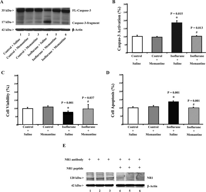
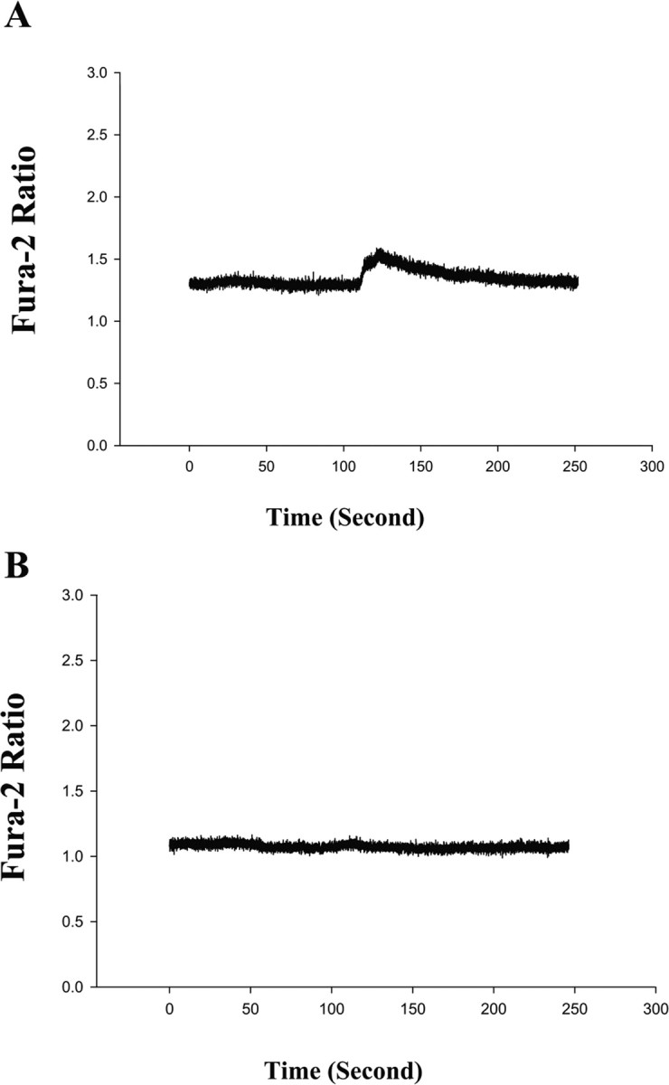
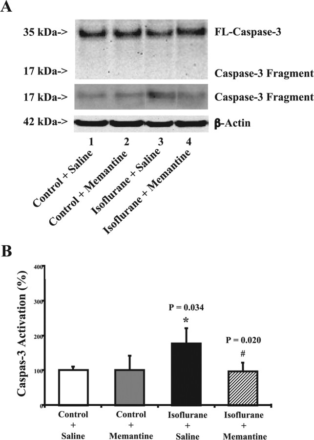
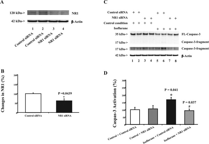
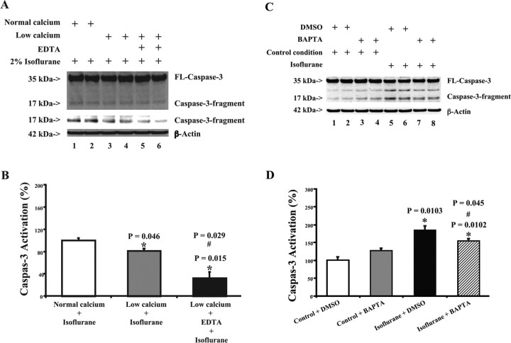
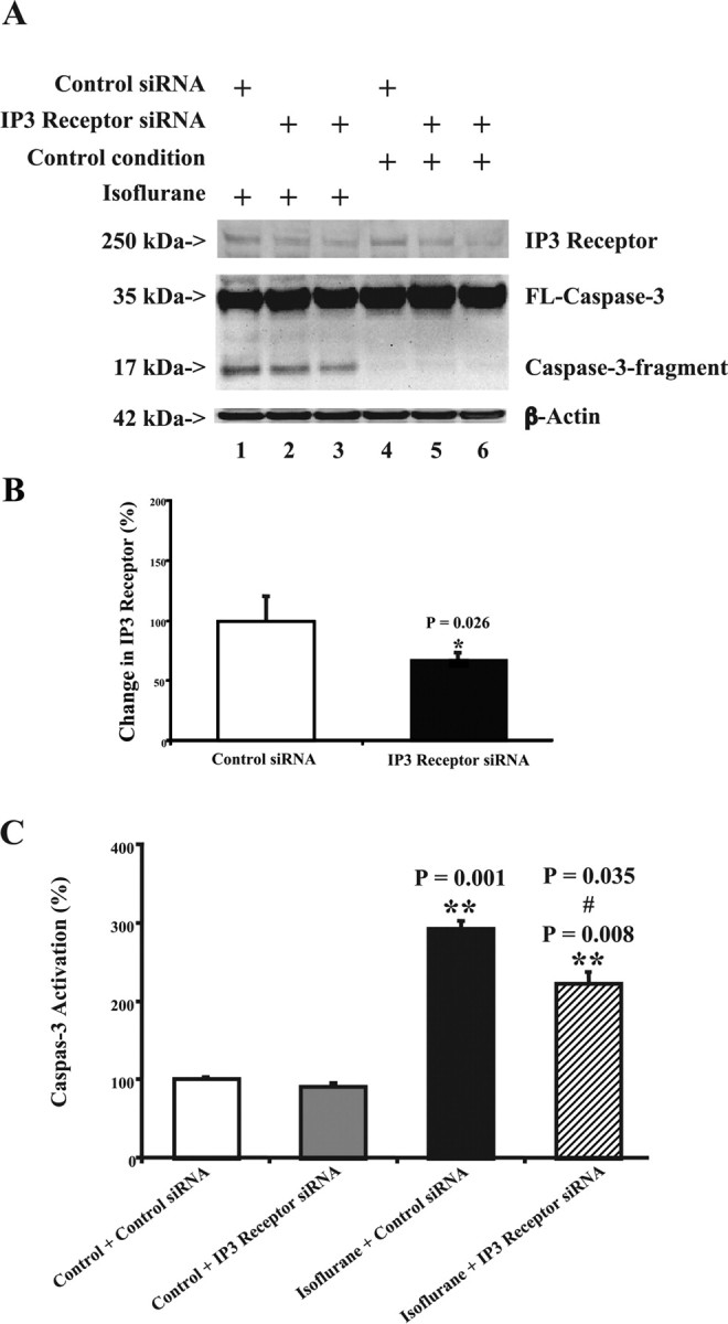
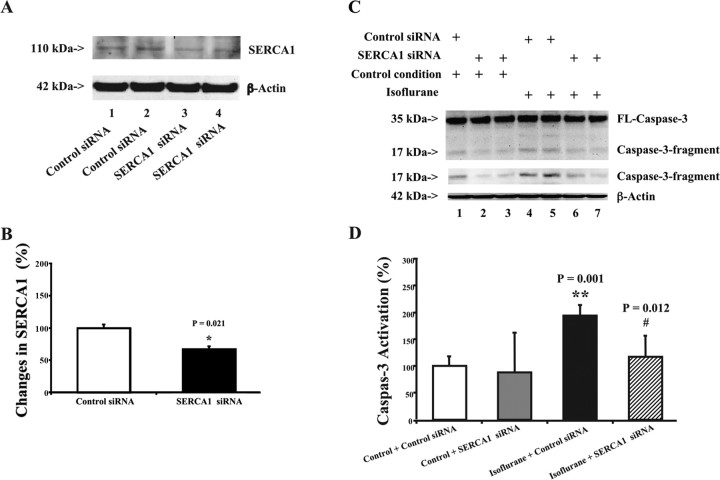
Increasing evidence indicates that caspase activation and apoptosis are associated with a variety of neurodegenerative disorders, including Alzheimer's disease. We reported that anesthetic isoflurane can induce apoptosis, alter processing of the amyloid precursor protein (APP), and increase amyloid-beta protein (Abeta) generation. However, the mechanism by which isoflurane induces apoptosis is primarily unknown. We therefore set out to assess effects of extracellular calcium concentration on isoflurane-induced caspase-3 activation in H4 human neuroglioma cells stably transfected to express human full-length APP (H4-APP cells). In addition, we tested effects of RNA interference (RNAi) silencing of IP(3) receptor, NMDA receptor, and endoplasmic reticulum (ER) calcium pump, sacro-/ER calcium ATPase (SERCA1). Finally, we examined the effects of the NMDA receptor partial antagonist, memantine, in H4-APP cells and brain tissue of naive mice. EDTA (10 mM), BAPTA (10 microM), and RNAi silencing of IP(3) receptor, NMDA receptor, or SERCA1 attenuated caspase-3 activation. Memantine (4 microM) inhibited isoflurane-induced elevations in cytosolic calcium levels and attenuated isoflurane-induced caspase-3 activation, apoptosis, and cell viability. Memantine (20 mg/kg, i.p.) reduced isoflurane-induced caspase-3 activation in brain tissue of naive mice. These results suggest that disruption of calcium homeostasis underlies isoflurane-induced caspase activation and apoptosis. We also show for the first time that the NMDA receptor partial antagonist, memantine, can prevent isoflurane-induced caspase-3 activation and apoptosis in vivo and in vitro. These findings, indicating that isoflurane-induced caspase activation and apoptosis are dependent on cytosolic calcium levels, should facilitate the provision of safer anesthesia care, especially for Alzheimer's disease and elderly patients.


Figures

Figure 1.
Figure 1.
Memantine attenuates isoflurane-induced caspase-3 activation and apoptosis in H4-APP cells. A, Treatment with 2% isoflurane for 6 h (lane 4) induces caspase-3 cleavage (activation) compared with control conditions (lane 1) in H4-APP cells. Treatment with 4 μm memantine alone does not cause caspase-3 activation (lanes 2 and 3) compared with control condition (lane 1). However, treatment with isoflurane plus memantine (lanes 5 and 6) causes a lesser degree of caspase-3 activation compared with isoflurane treatment alone (lane 4). There is no significant difference in amounts of β-actin in all of the above treatments. B, The 2% isoflurane treatment (black) increases caspase-3 activation compared with control conditions (white), normalized to β-actin levels. Memantine treatment alone (gray) does not induce caspase-3 activation compared with control condition (white); however, memantine treatment (lined) attenuates the isoflurane-induced (black) caspase-3 activation, normalized to β-actin levels. C, Isoflurane treatment (2%) (black) decreases cell viability compared with control conditions (white). Memantine treatment alone (gray) does not affect cell viability compared with control conditions (white); however, memantine treatment (lined) attenuates the isoflurane-induced reduction in cell viability (black). D, Isoflurane treatment (2%) (black) induces apoptosis compared with control conditions (white). Memantine treatment alone (gray) does not induce apoptosis compared with control conditions (white); however, memantine treatment (lined) attenuates isoflurane-induced apoptosis (black). E, H4-APP cells contain NR1. Western blot analysis illustrates that anti-NR1 antibody (lanes 1–3) detects NR1 in H4-APP cells, whereas NR1 peptide (lanes 4–6) prevents anti-NR1 antibody from detecting NR1 in H4-APP cells. These results suggest that there is NR1 in H4-APP cells. Data are means ± SD (n = 3–9 for each experimental group). A t test is used to compare the difference between control condition and the isoflurane or memantine treatment. The asterisk indicates the difference between isoflurane and control condition on caspase-3 activation (*p = 0.015), cell viability (*p = 0.001), and apoptosis (*p = 0.001); the difference between saline and memantine on isoflurane-induced caspase-3 activation (#p = 0.013), cell viability (#p = 0.037), and apoptosis (#p = 0.001) is indicated.
Figure 2.
Figure 2.
Isoflurane-induced elevation of cytosolic calcium levels can be inhibited by memantine in H4-APP cells. A, Isoflurane elevates cytosolic calcium levels in H4-APP cells. Isoflurane (1 mm; or 3.5%) was given to H4-APP cells at time 0. Cytosolic calcium levels were elevated after isoflurane treatment in H4-APP cells. B, Memantine inhibits isoflurane-induced elevation of cytosolic calcium levels in H4-APP cells. H4-APP cells were pretreated with 4 μm memantine for 5 min and then exposed to 1 mm (or 3.5%) isoflurane at time 0. Note that cytosolic calcium levels were not elevated by isoflurane in the memantine-pretreated H4-APP cells. These are representative tracings of two independent experiments.
Figure 3.
Figure 3.
Memantine attenuates isoflurane-induced caspase-3 activation in naive mice. A, Isoflurane treatment (lane 3) induces caspase-3 cleavage (activation) compared with control conditions (lane 1) in naive mice. Treatment with memantine alone does not cause caspase-3 activation (lane 2) compared with control condition (lane 1). However, treatment with isoflurane plus memantine (lanes 4) causes a reduction in caspase-3 activation compared with isoflurane treatment alone (lane 3). There is no significant difference in the amounts of β-actin in all of the treatments. The Western blot, which shows the caspase-3 fragment (17 kDa) only, is the same Western blot with longer exposure time during the film development. B, Isoflurane treatment (black) increases caspase-3 activation compared with control conditions (white), normalized to β-actin levels, in naive mice. Memantine treatment alone (gray) does not induce caspase-3 activation compared with control condition (white); however, memantine treatment (lined) attenuates the isoflurane-induced (black) caspase-3 activation, normalized to β-actin levels. Data are means ± SD (n = 4 for each experimental group). A t test is used to compare the difference between control condition and the isoflurane or memantine treatment. *p = 0.034, difference between isoflurane and control condition on caspase-3 activation; #p = 0.020, difference between saline and memantine on isoflurane-induced caspase-3 activation.
Figure 4.
Figure 4.
RNAi knockdown of NR1 reduces isoflurane-induced caspase-3 activation in H4-APP cells. A, NR1 siRNA (lanes 3 and 4) reduces protein levels of NR1 compared with control siRNA treatment (lanes 1 and 2). There is no significant difference in amounts of β-actin in control or NR1 siRNA-treated cells. B, Quantitation of the Western blots shows that NR1 siRNA treatment (black) reduces protein levels of NR1 compared with control siRNA treatment (white). A t test is used to compare difference between control siRNA and NR1 siRNA treatment in reducing NR1 protein levels (*p = 0.0439). C, Treatment with control siRNA plus isoflurane (lanes 5 and 6) induces caspase-3 activation compared with control siRNA plus control condition (lanes 1 and 2). NR1 siRNA alone (lanes 3 and 4) does not induce caspase-3 activation. However, treatment with NR1 siRNA plus isoflurane (lanes 7 and 8) induces a lesser degree of caspase-3 activation compared with a treatment with control siRNA plus isoflurane (lanes 5 and 6). The Western blot, which shows the caspase-3 fragment (17 kDa) only, is the same Western blot with longer exposure time during the film development. D, Quantification of the Western blot shows that control condition plus NR1 siRNA treatment (gray) dose not induce caspase-3 activation compared with control condition plus control siRNA treatment (white). The treatment with isoflurane plus control siRNA (black) induces caspase-3 activation compared with control condition plus control siRNA treatment (white). NR1 siRNA treatment (lined) attenuates the isoflurane-induced (black) caspase-3 activation. Data are means ± SD (n = 3–6 for each experimental group). A t test is used to compare the difference. *p = 0.041, difference between isoflurane and control condition on caspase-3 activation; #p = 0.037, difference between control siRNA and NR1 siRNA on isoflurane-induced caspase-3 activation.
Figure 5.
Figure 5.
EDTA, low calcium, and BAPTA attenuate isoflurane-induced caspase-3 activation in H4-APP cells. A, Treatment with isoflurane plus low-calcium condition (lanes 3 and 4) yields a reduction in caspase-3 cleavage (activation) compared with isoflurane treatment alone (lanes 1 and 2) in H4-APP cells. Treatment with isoflurane plus low-calcium condition and EDTA (lanes 5 and 6) causes an even lesser degree of caspase-3 activation compared with either isoflurane treatment alone (lanes 1 and 2) or the treatment of isoflurane plus low-calcium condition (lanes 3 and 4). There is no significant difference in the amounts of β-actin in all of the above treatments in H4-APP cells. The Western blot, which shows the caspase-3 fragment (17 kDa) only, is the same Western blot with longer exposure time during the film development. B, Caspase-3 activation assessed by quantifying the ratio of caspase-3 fragment to FL-caspase-3 in the Western blots. The treatment with isoflurane and low-calcium condition (gray) induces a reduction in caspase-3 activation compared with isoflurane treatment alone (white). The treatment with isoflurane plus low-calcium condition and EDTA (black) triggers an even lesser degree of caspase-3 activation compared with either isoflurane treatment alone (white) or the treatment with isoflurane plus low-calcium condition (gray). C, Treatment with DMSO plus isoflurane (lanes 5 and 6) induces caspase-3 activation compared with DMSO plus control condition (lanes 1 and 2). BAPTA alone (lanes 3 and 4) does not induce caspase-3 activation. However, treatment with BAPTA plus isoflurane (lanes 7 and 8) induces a lesser degree of caspase-3 activation compared with a treatment with DMSO plus isoflurane (lanes 5 and 6). D, Quantification of the Western blot shows that control condition plus BAPTA treatment (gray) dose not induce caspase-3 activation compared with control condition plus DMSO treatment (white). The treatment with isoflurane plus DMSO (black) induces caspase-3 activation compared with control condition plus DMSO treatment (white). BAPTA treatment (lined) attenuates the isoflurane-induced (black) caspase-3 activation. Data are means ± SD (n = 3–6 for each experimental group). A t test is used to compare the difference. *p = 0.046, difference between isoflurane plus low-calcium condition and isoflurane plus normal calcium condition; *p = 0.015, difference between isoflurane plus normal calcium condition and isoflurane plus low-calcium condition and EDTA; #p = 0.029, difference between isoflurane plus low-calcium condition and isoflurane plus low-calcium condition and EDTA; *p = 0.0103, difference between isoflurane and control condition on caspase-3 activation; #p = 0.045, difference between BAPTA and control treatment on isoflurane-induced caspase-3 activation.
Figure 6.
Figure 6.
RNAi knockdown of IP3 receptor reduces isoflurane-induced caspase-3 activation in H4-APP cells. A, IP3 receptor siRNA (lanes 2, 3, 5, and 6) reduces the protein levels of IP3 receptor compared with control siRNA treatment (lanes 1 and 4). The treatment with control siRNA plus isoflurane (lane 1) induces caspase-3 activation compared with control siRNA plus control condition (lane 4). The treatment of IP3 receptor siRNA plus control condition (lanes 5 and 6) does not induce caspase-3 activation compared with control siRNA plus control condition (lane 4). However, the treatment with IP3 receptor siRNA plus isoflurane (lanes 2 and 3) yields a reduction in caspase-3 activation compared with the treatment with control siRNA plus isoflurane (lane 1). B, Quantitation of the Western blots shows that IP3 receptor siRNA treatment (black) reduces the protein levels of IP3 receptor compared with control siRNA treatment (white). A t test is used to compare the difference between control siRNA and IP3 receptor siRNA treatment in reducing IP3 receptor protein levels (*p = 0.026). C, Control condition plus IP3 siRNA treatment (gray) dose not induce caspase-3 activation compared with control condition plus control siRNA treatment (white). The treatment with either isoflurane plus control siRNA (black) or isoflurane plus IP3 receptor siRNA (lined) causes caspase-3 activation compared with control condition plus control siRNA treatment (white). However, IP3 receptor siRNA treatment (lined) attenuates the isoflurane-induced (black) caspase-3 activation. Data are means ± SD (n = 3–6 for each experimental group). A t test is used to compare the difference. **p = 0.001, **p = 0.008, the difference between isoflurane and control condition on caspase-3 activation; #p = 0.035, the difference between control siRNA and IP3 receptor siRNA on the isoflurane-induced caspase-3 activation.
Figure 7.
Figure 7.
RNAi knockdown of SERCA1 reduces isoflurane-induced caspase-3 activation in H4-APP cells. A, SERCA1 siRNA (lanes 3 and 4) reduces protein levels of SERCA1 compared with control siRNA treatment (lanes 1 and 2). There is no significant difference in amounts of β-actin in control or SERCA1 siRNA-treated cells. B, Quantitation of the Western blots shows that SERCA1 siRNA treatment (black) reduces protein levels of SERCA1 compared with control siRNA treatment (white). A t test is used to compare the difference between control siRNA and SERCA1 siRNA treatment in reducing SERCA1 protein levels (*p = 0.021). C, Treatment with control siRNA plus isoflurane (lanes 4 and 5) induces caspase-3 activation compared with control siRNA plus control condition (lane 1). SERCA1 siRNA alone (lanes 2 and 3) does not induce caspase-3 activation. However, treatment with SERCA1 siRNA plus isoflurane (lanes 6 and 7) induces a reduction in caspase-3 activation compared with the treatment with control siRNA plus isoflurane (lanes 4 and 5). The Western blot, which shows the caspase-3 fragment (17 kDa) only, is the same Western blot with longer exposure time during the film development. D, Quantification of the Western blot shows that control condition plus SERCA1 siRNA treatment (gray) dose not induce caspase-3 activation compared with control condition plus control siRNA treatment (white). Treatment with isoflurane plus control siRNA (black) induces caspase-3 activation compared with control condition plus control siRNA treatment (white). SERCA1 siRNA treatment (lined) attenuates the isoflurane-induced caspase-3 activation (black). Data are means ± SD (n = 4 for each experimental group). A t test is used to compare the difference. **p = 0.001, difference between isoflurane and control condition on caspase-3 activation; #p = 0.012, difference between control siRNA and SERCA1 siRNA on isoflurane-induced caspase-3 activation.

References

    1. Berridge MJ. Inositol trisphosphate and calcium signalling. Nature. 1993;361:315–325. - PubMed
    1. Bickler PE, Fahlman CS. The inhaled anesthetic, isoflurane, enhances Ca2+-dependent survival signaling in cortical neurons and modulates MAP kinases, apoptosis proteins and transcription factors during hypoxia. Anesth Analg. 2006;103:419–429. table of contents. - PubMed
    1. Bickler PE, Zhan X, Fahlman CS. Isoflurane preconditions hippocampal neurons against oxygen-glucose deprivation: role of intracellular Ca2+ and mitogen-activated protein kinase signaling. Anesthesiology. 2005;103:532–539. - PubMed
    1. Bohnen N, Warner MA, Kokmen E, Kurland LT. Early and midlife exposure to anesthesia and age of onset of Alzheimer's disease. Int J Neurosci. 1994a;77:181–185. - PubMed
    1. Bohnen NI, Warner MA, Kokmen E, Beard CM, Kurland LT. Alzheimer's disease and cumulative exposure to anesthesia: a case-control study. J Am Geriatr Soc. 1994b;42:198–201. - PubMed

Publication types