. 2008 Apr 29;105(17):6243-8.
doi: 10.1073/pnas.0801569105. Epub 2008 Apr 23.

Molecular architecture of Galphao and the structural basis for RGS16-mediated deactivation

Affiliations

Molecular architecture of Galphao and the structural basis for RGS16-mediated deactivation

Kevin C Slep et al. Proc Natl Acad Sci U S A. .

Abstract

Heterotrimeric G proteins relay extracellular cues from heptahelical transmembrane receptors to downstream effector molecules. Composed of an alpha subunit with intrinsic GTPase activity and a betagamma heterodimer, the trimeric complex dissociates upon receptor-mediated nucleotide exchange on the alpha subunit, enabling each component to engage downstream effector targets for either activation or inhibition as dictated in a particular pathway. To mitigate excessive effector engagement and concomitant signal transmission, the Galpha subunit's intrinsic activation timer (the rate of GTP hydrolysis) is regulated spatially and temporally by a class of GTPase accelerating proteins (GAPs) known as the regulator of G protein signaling (RGS) family. The array of G protein-coupled receptors, Galpha subunits, RGS proteins and downstream effectors in mammalian systems is vast. Understanding the molecular determinants of specificity is critical for a comprehensive mapping of the G protein system. Here, we present the 2.9 A crystal structure of the enigmatic, neuronal G protein Galpha(o) in the GTP hydrolytic transition state, complexed with RGS16. Comparison with the 1.89 A structure of apo-RGS16, also presented here, reveals plasticity upon Galpha(o) binding, the determinants for GAP activity, and the structurally unique features of Galpha(o) that likely distinguish it physiologically from other members of the larger Galpha(i) family, affording insight to receptor, GAP and effector specificity.


Conflict of interest statement

The authors declare no conflict of interest.

Figures

Fig. 1.
Fig. 1.
Structure of the Gαo·GDP·AlF4·RGS16 complex. The Gαo·GDP·AlF4·RGS16 complex is represented in ribbons format. RGS16 is indicated in gray; Gαo GTPase domain is in green with nucleotide-dependent switch regions highlighted in orange; Gαo helical domain is in purple. Structure at Right is rotated 90° about the y axis relative to the structure at Left.
Fig. 2.
Fig. 2.
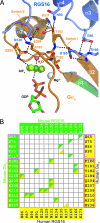
o–RGS16 contacts and the RGS domain GAP mechanism. (A) Stick-and-ribbons diagram of the Gαo GTP binding pocket occupied by the transition state analog of GTP hydrolysis; GDP·AlF4 is shown with Mg2+ and the attacking water. Gαo is shown in green and orange (switch regions). RGS16 is shown in purple. RGS16 residues do not contact the GTP or attacking water directly; instead they buttress the endogenous catalytic residues of Gαo, stabilizing their conformation in the transition state. (B) Comparative <4 Å electrostatic interaction matrix between RGS16 and Gα subunits. Electrostatic interactions between mouse Gαo and mouse RGS16 are indicated in green. Electrostatic interactions between human Gαi1 and human RGS16 are indicated in yellow (PDB ID code 2IK8; see ref. 18). Gα switch residues are boxed in orange; helical domain residues are boxed in purple. Interactions that occur in one or both crystallographic protomers are included for both Gαo–RGS16 and Gαi1–RGS16.
Fig. 3.
Fig. 3.
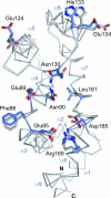
Determinants of RGS16 binding and conformational plasticity. Structural alignment of mouse RGS16 in the free (gray) and Gαo-bound (slate) states. The Cα trace is presented for both structures, with key residues used in the Gαo interaction represented in stick format. The structural alignment was performed with PyMol.
Fig. 4.
Fig. 4.
Unique structural determinants in the Gαo helical domain. (A) Structural alignment of Gαo and Gαi1 bound to GDP·AlF4, coordinates taken from their respective complex structures with RGS16 and RGS4 [PDB ID code 1AGR (5)] and displayed as a Cα trace. Gαo is colored as in Fig. 1A except that the divergent αB–αC loop is shown in red. Gαi1 is shown in gray except for the comparative αB–αC loop, which is shown in blue. The structural alignment was performed with PyMol. (B) Structural alignment as in A, zoomed in on the αB–αC loop region. Gαi/12 [PDB ID code 1ZCA (36)] is included, with its respective αBd–αC loop region shown in yellow. (C) Representation of Gαo in ribbon format (Left, colored as in Fig. 1A) for orientation and in CPK (Right) with the GTPase domain, helical domain, and switch regions colored white, light gray, and dark gray respectively. Residues identical across Gαi1-3 and Gαt but not identical in Gαo are mapped in red. Residues identical across Gαi1-3 but not identical in Gαo are mapped in blue. Two orientations, related by 180° rotations, are displayed vertically.

References

    1. Chan RK, Otte CA. Isolation and genetic analysis of Saccharomyces cerevisiae mutants supersensitive to G1 arrest by a factor and alpha factor pheromones. Mol Cell Biol. 1982;2:11–20. - PMC - PubMed
    1. Koelle MR, Horvitz HR. EGL-10 regulates G protein signaling in the C. elegans nervous system and shares a conserved domain with many mammalian proteins. Cell. 1996;84:115–125. - PubMed
    1. Berman DM, Wilkie TM, Gilman AG. GAIP and RGS4 are GTPase-activating proteins for the Gi subfamily of G protein alpha subunits. Cell. 1996;86:445–452. - PubMed
    1. Berman DM, Kozasa T, Gilman AG. The GTPase-activating protein RGS4 stabilizes the transition state for nucleotide hydrolysis. J Biol Chem. 1996;271:27209–27212. - PubMed
    1. Tesmer JJ, Berman DM, Gilman AG, Sprang SR. Structure of RGS4 bound to AlF4-activated G(i alpha1): Stabilization of the transition state for GTP hydrolysis. Cell. 1997;89:251–261. - PubMed

Publication types