. 2008 Apr 29;105(17):6421-6.
doi: 10.1073/pnas.0801383105. Epub 2008 Apr 23.

Unusually rapid evolution of Neuroligin-4 in mice

Affiliations

Unusually rapid evolution of Neuroligin-4 in mice

Marc F Bolliger et al. Proc Natl Acad Sci U S A. .

Abstract

Neuroligins (NLs) are postsynaptic cell-adhesion molecules that are implicated in humans in autism spectrum disorders because the genes encoding NL3 and NL4 are mutated in rare cases of familial autism. NLs are highly conserved evolutionarily, except that no NL4 was detected in the currently available mouse genome sequence assemblies. We now demonstrate that mice express a distant NL4 variant that rapidly evolved from other mammalian NL4 genes and that exhibits sequence variations even between different mouse strains. Despite its divergence, mouse NL4 binds neurexins and is transported into dendritic spines, suggesting that the core properties of NLs are retained in this divergent NL isoform. The selectively rapid evolution of NL4 in mice suggests that its function in the brain is under less stringent control than that of other NLs, shedding light on why its mutation in autism spectrum disorder patients is not lethal, but instead leads to a discrete developmental brain disorder.


Conflict of interest statement

The authors declare no conflict of interest.

Figures

Fig. 1.
Fig. 1.
Sequence variations in NL4* from different mouse strains. (A) The complete coding sequence of NL4* was amplified by PCR on brain cDNA prepared from a pool of 1,000 BALB/c mice and was cloned and sequenced. A total of 26 clones were analyzed and found to comprise 10 cDNA variants that feature 18 nt substitutions (circles) and four insertions/deletions (rectangles) as listed. The bar shows schematically the mRNA of NL4* with the location of exons in the NL4* gene (TM, transmembrane region; ATG, initiator codon; TAG, stop codon). See Table 2 for a list of the changes observed. (B) Exons 1, 6, and 7 were PCR-amplified on genomic DNA isolated from three mouse strains (arrows indicate the location of PCR primers). The sequences obtained from BALB/c and C57BL/6 mice were identical, whereas the strain 129/Sv sequence exhibited eight changes in exon 6 and two changes in exon 7, with one of the changes leading to an amino acid substitution. See also Table 3.
Fig. 2.
Fig. 2.
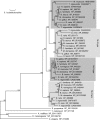
Phylogenetic analysis of NL sequences. Vertebrate NLs form four groups according to the four major types of NLs. Possible orthologs identified in invertebrates (insects, sea urchin, and C. elegans) serve as outgroup sequences and determine the root of the four groups of vertebrate NLs. The tree was built by using the MOLPHY software, and sequences are labeled with species names and GenBank accession numbers. Local bootstrap values are indicated (as percentages) at internal tree nodes.
Fig. 3.
Fig. 3.
Organization and size of NL genes. The contribution of the seven coding exons to the four mouse NLs and human NL4 and NL5 proteins is shown (TM, transmembrane region). For simplification, the exon containing the translational start codon is denoted as exon 1, and the exon containing the stop codon is denoted as exon 7 in all NL genes. Most, if not all, NL genes contain at least one 5′ noncoding exon not shown. Exon 2 encoding splice insert A1 is present only in NL1 and NL3, whereas exon 3 (splice insert A2) can be found in all NL genes. Only the NL1 gene is alternatively spliced at the end of exon 5 to create splice site B. Numbers inserted between exons give the size of corresponding introns in kilobases. Numbers on the left denote gene sizes (as measured from the translational start codon in exon 1 to the stop codon in exon 7). n.a., not available.
Fig. 4.
Fig. 4.
Organization of the mouse NL4* gene. (A) Schematic diagram of the mouse NL4* gene structure as determined by sequencing of genomic DNA (GenBank accession number EU350930). The gene contains six coding exons and at least one noncoding (exon 0). Exons are indicated by filled boxes and are numbered. Exon 3 encodes the alternative spliced A2 insert, whereas no exon corresponding to exon 2 in NL1 and NL3 (which encodes the alternatively spliced A1 insert) is present. Sequence analyses identified five repetitive DNA regions (R1, R2, S1, S2, and T; open boxes), the first four of which are closely related to each other as shown in the sequence alignment in B. The copy numbers and consensus sequences of the repetitive regions (determined with a cut-off of 80%) are indicated (P, location of 681-bp hybridization probe used to isolate the genomic DNA clone). (B) Sequence alignment of the repetitive regions R1, R2, S1, and S2 with identical bases boxed on a black background; note that the four repetitive regions form two pairs that are present in opposite orientations.
Fig. 5.
Fig. 5.
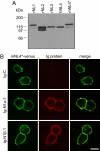
Expression of NL4* in nonneuronal cells and binding to neurexins. (A) COS cells were transiently transfected with expression vectors encoding rat NL1-NL3, human NL4, or mouse NL4*. Protein extracts were analyzed by SDS/PAGE and immunoblotting using the pan-NL antibody 19C. Molecular-mass markers (kDa) are shown on the left. (B) HEK-293T cells expressing venus-tagged mouse NL4* (green) were incubated with soluble Ig-neurexin or Ig-control proteins. Cell surface-bound Ig-protein was visualized with fluorescent secondary antibodies (red). This binding assay revealed that NL4* binds both α-neurexin (Ig-N1α-1) and β-neurexin (Ig-N1β-1) but not the Ig-control protein (Ig-C). The images on the right show the merged pictures of neurexin and NL stainings with coincident labeling shown in yellow (Scale bar, 10 μm.)
Fig. 6.
Fig. 6.
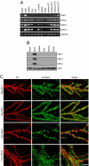
Tissue expression of NL isoforms and localization in transfected neurons. (A) RT-PCR analysis of NLs with primers specific for NL1, NL2, NL3, NL4*, or glyceraldehyde-3-phosphate dehydrogenase (G3PDH, control) was performed on cDNA preparations made from mouse tissue RNA as listed. Products were resolved on 1% agarose gels. (B) Immunoblot analysis of NLs in mouse tissues by using isoform-specific antibodies 4C12 (NL1), 169C (NL2), and 639B (NL3). NLs could be detected exclusively in the brain homogenate. Valosin-containing protein (VCP, antibody 443B) served as a loading control. (C) Dissociated hippocampal neurons isolated from newborn rat pups were transfected with vectors encoding flag-tagged rat NL1 or NL2, myc-tagged human NL4, or myc-tagged mouse NL4* on DIV 10. Six days later, cultures were fixed and stained with immunofluorescent antibodies to the flag or myc epitopes (red) and synapsin (green; antibody E028). All four NL isoforms localized to dendritic spines, whereas the synapsin immunoreactivity was observed presynaptically (Scale bar, 10 μm.)

Similar articles

Cited by

References

    1. Ichtchenko K, et al. Neuroligin 1: A splice site-specific ligand for β-neurexins. Cell. 1995;81:435–443. - PubMed
    1. Ichtchenko K, Nguyen T, Südhof TC. Structures, alternative splicing, and neurexin binding of multiple neuroligins. J Biol Chem. 1996;271:2676–2682. - PubMed
    1. Ushkaryov YA, Petrenko AG, Geppert M, Südhof TC. Neurexins: Synaptic cell surface proteins related to the α-latrotoxin receptor and laminin. Science. 1992;257:50–56. - PubMed
    1. Nguyen T, Südhof TC. Binding properties of neuroligin 1 and neurexin 1β reveal function as heterophilic cell adhesion molecules. J Biol Chem. 1997;272:26032–26039. - PubMed
    1. Scheiffele P, Fan J, Choih J, Fetter R, Serafini T. Neuroligin expressed in nonneuronal cells triggers presynaptic development in contacting axons. Cell. 2000;101:657–669. - PubMed

Publication types

MeSH terms

Associated data