Review
. 2008 May;87(5):414-34.
doi: 10.1177/154405910808700509.

Mechanisms of tooth eruption and orthodontic tooth movement

Affiliations
Review

Mechanisms of tooth eruption and orthodontic tooth movement

G E Wise et al. J Dent Res. 2008 May.

Abstract

Teeth move through alveolar bone, whether through the normal process of tooth eruption or by strains generated by orthodontic appliances. Both eruption and orthodontics accomplish this feat through similar fundamental biological processes, osteoclastogenesis and osteogenesis, but there are differences that make their mechanisms unique. A better appreciation of the molecular and cellular events that regulate osteoclastogenesis and osteogenesis in eruption and orthodontics is not only central to our understanding of how these processes occur, but also is needed for ultimate development of the means to control them. Possible future studies in these areas are also discussed, with particular emphasis on translation of fundamental knowledge to improve dental treatments.
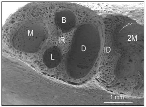

Figures

Figure 1
Figure 1
Comparison of compression and tension zones. (A) SEM of resorption cratering in a rat maxillary first molar root adjacent to a compression zone with 40 cN of orthodontic force for 5 days. (B) SEM of a rat maxillary first molar socket. Osteogenesis in response to 40 cN of orthodontic tension for 5 days can be seen as bony spicules extending into the socket from the top of the image.
Figure 2
Figure 2
Morphology of unerupted tooth vs. erupted tooth. (A) Longitudinal section of a rat first mandibular molar at day 3 postnatally, showing the loose connective tissue sac, the dental follicle (DF), that surrounds the 1st molar (M1). Note that the unerupted tooth is encased in alveolar bone (AB), and that a prominent stellate reticulum is present. (B) Fluorescence micrograph image of a rat maxillary first molar and adjacent tissue with 40 cN of orthodontic force for 5 days. Root (R), periodontal ligament (P), and alveolar bone at compression (C) and tension sites (T). Tetracycline bone markers (orange and yellow) have been incorporated into the alveolar bone to demonstrate the bone turnover patterns. Note that the tension side is largely osteogenic, while the compression side is marked by cratering of both the bone and root, indicating resorption; however, some of the craters are also osteogenic, indicating that remodeling is taking place.
Figure 3
Figure 3
Graph depicting the expression of genes in the dental follicle of the rat 1st mandibular molar at various days post-natally. At day 3, the major burst of osteoclastogenesis, osteoprotegerin is down-regulated such that a favorable RANK/osteoprotegerin ratio is established to promote osteoclastogenesis. At this time, CSF-1 is maximally expressed, both to down-regulate osteoprotegerin expression and to promote osteoclast precursor recruitment and osteoclastogenesis. At day 10, the minor burst of osteoclastogenesis, VEGF is maximally expressed to stimulate RANK expression on osteoclast precursors, as well as to interact with RANKL and CSF-1 to promote osteoclastogenesis. At this time, RANKL is up-regulated such that a favorable RANKL/osteoprotegerin ratio could exist to stimulate this minor burst of osteoclastogenesis.
Figure 4
Figure 4
High-power SEM view of a portion of the wall of the alveolar bony crypt of a 1st mandibular rat molar at day 3 post-natally, extending from the coronal region (C) of the wall to the basal region (B). Although only a small area of the large coronal region is shown, note elongated depressions (D) that give a scalloped appearance to the bone in that region, whereas in the basal region the bone consists of many narrow trabeculae (T). Scalloped bone is undergoing resorption, whereas trabecular bone reflects bone formation. Between the two is an intermediate region (I) of bone that is relatively smooth. Such smooth bone is more inert, or neutral, reflecting neither growth nor resorption.
Figure 5
Figure 5
Low-power SEM view of the alveolar bony crypt of the rat 1st mandibular molar at day 14, showing the extensive bone growth that has occurred at the base, especially in the region of the interradicular septum (IR). Four pits represent where the 4 roots reside—mesial (M), distal (D), mesiolabial (L), and mesiobuccal (B). A prominent interdental septum (ID) is present that separates the crypt of the first molar from the 2nd molar (2M). Note that all the newly formed bone, such as seen in the IR or ID, is trabecular.

References

    1. Adachi T, Sato K, Tomita Y. Directional dependence of osteoblastic calcium response to mechanical stimuli. Biomech Model Mechanobiol. 2003;2:73–82. - PubMed
    1. Ajubi NE, Klein-Nulend J, Alblas MJ, Burger EH, Nijweide PJ. Signal transduction pathways involved in fluid flow-induced PGE2 production by cultured osteocytes. Am J Physiol. 1999;276:171–178. - PubMed
    1. Alhashimi N, Frithiof L, Brudvik P, Bakhiet M. Orthodontic tooth movement and de novo synthesis of proinflammatory cytokines. Am J Orthod Dentofacial Orthop. 2001;119:307–312. - PubMed
    1. Anderson DM, Maraskovsky E, Billingsley WL, Dougall WC, Tometsko ME, Roux ER, et al. A homologue of the TNF receptor and its ligand enhance T-cell growth and dendritic-cell function. Nature. 1997;390:175–179. - PubMed
    1. Apajalahti S, Sorsa T, Railavo S, Ingman T. The in vivo levels of matrix metalloproteinase-1 and -8 in gingival crevicular fluid during initial orthodontic tooth movement. J Dent Res. 2003;82:1018–1022. - PubMed

Publication types

MeSH terms

LinkOut - more resources