. 2008 Jul;19(7):2830-43.
doi: 10.1091/mbc.e07-12-1236. Epub 2008 Apr 23.

Coat-tether interaction in Golgi organization

Affiliations

Coat-tether interaction in Golgi organization

Yusong Guo et al. Mol Biol Cell. 2008 Jul.

Abstract

Biogenesis of the Golgi apparatus is likely mediated by the COPI vesicle coat complex, but the mechanism is poorly understood. Modeling of the COPI subunit betaCOP based on the clathrin adaptor AP2 suggested that the betaCOP C terminus forms an appendage domain with a conserved FW binding pocket motif. On gene replacement after knockdown, versions of betaCOP with a mutated FW motif or flanking basic residues yielded a defect in Golgi organization reminiscent of that occurring in the absence of the vesicle tether p115. Indeed, betaCOP bound p115, and this depended on the betaCOP FW motif. Furthermore, the interaction depended on E(19)E(21) in the p115 head domain and inverse charge substitution blocked Golgi biogenesis in intact cells. Finally, Golgi assembly in permeabilized cells was significantly reduced by inhibitors containing intact, but not mutated, betaCOP FW or p115 EE motifs. Thus, Golgi organization depends on mutually interacting domains in betaCOP and p115, suggesting that vesicle tethering at the Golgi involves p115 binding to the COPI coat.


Figures

Figure 1.
Figure 1.
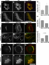
βCOP knockdown. (A) HeLa cells expressing GalNAcT2-GFP, grown in 35-mm dishes also containing a coverslip, were either mock transfected or transfected with βCOP siRNA. Total cell extracts (20 μg/lane) were analyzed by immunoblotting by using anti-βCOP, and, as a loading control, anti-p115 antibodies. The coverslips were analyzed by immunofluorescence by using anti-βCOP antibody. Quantified βCOP levels determined by immunoblot (Blot: mean βCOP/p115 ± SD; n = 2) and immunofluorescence (IF: mean ± SEM; >15 cells each) are presented after normalization to the mock-transfected cells. (B–J) HeLa cells expressing GalNAcT2-GFP were mock transfected (B–D) or transfected with either of two siRNAs against βCOP (E–J). The GalNAcT2-GFP, giantin (Gtn) and βCOP distributions were visualized 48 h later. Bar, 10 μm. (K–N) HeLa cells expressing GalNAcT2-GFP were mock transfected (K and L) or transfected with siRNA against βCOP (M and N) and GalNAcT2-GFP and eCOP distributions were visualized 48 h later. Bar, 10 μm.
Figure 2.
Figure 2.
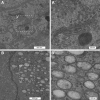
Absence of Golgi stacks. Mock (A) and siRNA (B) transfected GalNAcT2-GFP HeLa cells were processed for electron microscopy. Outlined regions are shown at higher magnifications in the adjacent panels as indicated (A′ and B′). Note the striking juxtanuclear accumulation of aggregated membranes exhibiting diameters of ∼250 nm in βCOP knockdown cells.
Figure 3.
Figure 3.
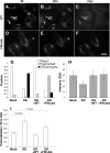
Analysis of Golgi remnant structures after βCOP knockdown. (A–F) Mock- (A–C) or βCOP siRNA (D–F)-treated cells were processed to reveal GalNAcT2-GFP and giantin (Gtn) staining in juxtanuclear Golgi/remnants with 3D rendering. The merged images have GalNAcT2-GFP in green and giantin in red. Bar, 5 μm. (G) Golgi/remnant fluorescent size was determined in mock and βCOP knockdown cells (mean ± SD; >15 cells each). Size was total volume occupied by above-threshold fluorescence of giantin and showed a significant increase in βCOP knockdown cells. (H) Quantified giantin staining intensity for mock and βCOP knockdown cells is shown (n = 2, mean ± SD; >15 cells/condition/experiment). Intensity is the normalized mean pixel value in the Golgi/remnant. (I–N) HeLa cells stably expressing GalNAcT2-GFP were either mock (I–K) or βCOP siRNA (L–N) transfected, and, after 24 h, retransfected with a plasmid encoding KDEL-RFP. After another 24-h incubation, the cells were processed to reveal GalNAcT2-GFP and KDEL-RFP. The merged images have GalNAcT2-GFP in green and KDEL-RFP in red. Note the accumulation of KDEL-RFP in the juxtanuclear remnants induced by βCOP knockdown (arrows). (O–U) Mock- (O–Q) and βCOP (R–T) siRNA-transfected cells were costained using immunofluorescence to compare colocalization between giantin and ERGIC53. Colocalization was quantified (U) by determining the average of fraction of each marker's area that was also occupied by the other marker, where area is determined by above threshold fluorescence and corresponds to a Golgi/remnant structure (n = 2, mean ± SD; >15 cells each). Bar, 10 μm.
Figure 4.
Figure 4.
Computational model of βCOP C terminus. (A–C) Shown are the determined structures for the C-termini of αAP2 and γCOP and the computed model for the analogous C terminus of βCOP. The βCOP model was generated using ModBase (Pieper et al., 2006) and based on βCOP sequence homology to γCOP. The position of the conserved FW residues is indicated in red. They lie on a surface exposed α-helix of the appendage domain, and they are flanked by three basic residues shown in green. (D) Schematic diagram of the COPI complex based on homology to AP2 (modified from Hoffman et al., 2003). (E) Schematic diagram of the βCOP constructs used in this study (numbering refers to amino acids).
Figure 5.
Figure 5.
βCOP appendage FW motif is required for Golgi biogenesis. (A–F) βCOP siRNA-transfected cells were retransfected after 24 h with plasmid encoding myc-tagged rat βCOP WT (A–C) or the FW>AA mutant (D–F). After an additional 24 h the cells were processed to assess Golgi integrity, by using GalNAcT2-GFP and giantin (Gtn) staining, and construct expression, by using myc-epitope staining. (G) Golgi integrity in mock, knockdown (KD) cells, and myc-positive knockdown cells (KD+WT or KD+FW>AA) was then scored as the fraction of cells showing normal Golgi ribbons (Ribbon), dispersed punctate structures (Fragmented), and juxtanuclear aggregated structure accompanied by partially ER-localized GalNAcT2-GFP (ER+remnants). Note that wild-type βCOP restored ribbon structure, whereas βCOP FW>AA yielded an IC pattern. (H–I) Quantification of giantin fluorescence intensity and giantin versus GalNAcT2-GFP colocalization was carried as described in the Figure 3 legend (mean ± SD; >15 cells each). Whereas wild-type βCOP restored marker intensity and separation, βCOP FW>AA only restored intensity.
Figure 6.
Figure 6.
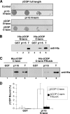
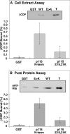
βCOP FW motif binds p115. (A) Yeast two-hybrid assay growth results for full-length βCOP (as bait) tested against a vector only control and the p115 N terminus. The p115 N terminus (as bait) was also tested against the βCOP N-terminal and C-terminal constructs diagrammed in Figure 4E. (B) Equivalent amounts (≈5 μg) of bead-bound GST and GST-p115-N were incubated with 1.5 μg of purified hexahistidine-tagged βCOP N-terminal and C-terminal constructs diagrammed in Figure 4E. Recovery of the βCOP constructs was determined by immunoblotting with an anti-histidine epitope antibody and compared with the 0.15 μg of signal representing 10% of total (T). (C) Equivalent amounts (≈5 μg) of bead-bound GST and GST-p115-N were incubated with 1.5 μg of purified hexahistidine-tagged βCOP C-terminal constructs with and without the FW>AA mutation as diagrammed in Figure 4E. Recovery of the βCOP constructs was determined by immunoblotting with an anti-histidine epitope antibody and compared with the 0.075-μg signal representing 5% of total (T). (D) The results showing a requirement for the FW motif in the βCOP–p115 interaction were quantified to yield the bound βCOP as a percentage of total (mean ± SD; n = 2).
Figure 7.
Figure 7.
p115 E19E21 binds βCOP. (A) Equivalent amounts (≈5 μg) of bead-bound GST, GST-p115-N, and GST-p115-N with the E19,21K mutation were incubated with rabbit liver cytosol. Recovery of bound βCOP was determined by immunoblotting. Quantification presents the fraction bound as a percentage of total (mean ± SD; n = 3). (B) The purified hexahistidine-tagged βCOP C terminus (1.5 μg) was incubated with glutathione beads bearing ≈5 μg of GST, GST-p115-N, or GST-p115-N with the E19,21K mutation. Bound protein was determined by immunoblotting with the anti-histidine epitope antibody and compared with 0.075-μg signal representing 5% of total. The bound as a percentage of total is shown (mean ± SD; n = 3).
Figure 8.
Figure 8.
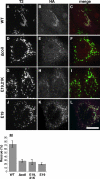
p115 E19E21 is required for Golgi biogenesis. (A–L) GalNAcT2-GFP expressing HeLa cells were transfected with p115 siRNA and after 72 h, the cells were microinjected with plasmids encoding hemagglutinin (HA)-tagged bovine p115 with sequences corresponding to wild-type (A–C), Δcoil1 (D–F), E19,21K (G–I), or E19K (J–L). After 5 h, the cells were fixed and stained with anti-HA antibody to detect microinjected cells, and GalNAcT2-GFP was visualized to determine Golgi integrity. (M) The results were quantified by determining percentage of HA-epitope–positive cells with an intact Golgi (n = 3, mean ± SD; >15 cells/experiment/condition).
Figure 9.
Figure 9.
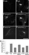
In vitro Golgi assembly. (A–F) NRK cells were sequentially treated with BFA and H89 to collapse the Golgi into the ER, permeabilized with streptolysin-O, washed with salt solution, and incubated in reaction mix for 50 min at 37°C. The reaction mix contained buffer, 40 μg/ml GFP, and an ATP-regenerating system in the absence (A) or presence of cytosol (B) supplemented with either 0.2 mg/ml purified βCOP-C (C), 0.2 mg/ml βCOP-CFW>AA (D), 0.1 mg/ml p115-N (E), or 0.1 mg/ml p115-NE19,21K (F). After incubation, the cells were fixed and stained with anti-giantin antibody to assay Golgi assembly, and GFP was visualized to confirm permeabilization. (G) The results were quantified by determining percentage of GFP-positive cells with a juxtanuclear Golgi (n = 2; mean ± SD; >15 cells/experiment/condition).
Figure 10.
Figure 10.
Model depicting COPIER and COPIG pathways. (A) Two COPI retrieval pathways are diagrammed. The COPIER pathway, which is p115-independent, functions to concentrate Golgi proteins via ER retrieval of non-Golgi proteins. The COPIG pathway, which is dependent on p115 binding to βCOP, functions to compartmentalize Golgi proteins via local retrieval between Golgi cisternae and from the Golgi to the IC. (B) In βCOP knockdown cells, both COPIER and COPIG are blocked, but COPII vesicle formation and fusion continues. This induces accumulation of pre-ICs contaminated with ER resident proteins and other proteins that fail to recycle. In the absence of recycling, Golgi proteins fail to concentrate. (C) Inhibition of the p115-βCOP interaction leaves the COPIG pathway blocked, whereas COPIER functions. Retrieval by COPIER allows concentration of Golgi proteins in ICs, but the membranes fail to compartmentalize without cycles of COPIG retrieval. (D) A hypothetical COPIG sorting and tethering complex is diagramed. Recruitment of p115 into COPI vesicles occurs when the E19E21 motif in p115 head domain binds the βCOP appendage FW domain. Before vesicle uncoating, this interaction could also dock the vesicles when the p115 coiled-coil domain simultaneously binds a SNARE complex on the target membrane.

Comment in

  • Mol Biol Cell. 19:2681.

Similar articles

Cited by

References

    1. Allan B. B., Moyer B. D., Balch W. E. Rab1 recruitment of p115 into a cis-SNARE complex: programming budding COPII vesicles for fusion. Science. 2000;289:444–448. - PubMed
    1. Alvarez C., Fujita H., Hubbard A., Sztul E. ER to Golgi transport: requirement for p115 at a pre-Golgi VTC stage. J. Cell Biol. 1999;147:1205–1222. - PMC - PubMed
    1. Andag U., Neumann T., Schmitt H. D. The coatomer-interacting protein Dsl1p is required for Golgi-to-endoplasmic reticulum retrieval in yeast. J. Biol. Chem. 2001;276:39150–39160. - PubMed
    1. Andag U., Schmitt H. D. Dsl1p, an essential component of the Golgi-endoplasmic reticulum retrieval system in yeast, uses the same sequence motif to interact with different subunits of the COPI vesicle coat. J. Biol. Chem. 2003;278:51722–51734. - PubMed
    1. Appenzeller-Herzog C., Hauri H. P. The ER-Golgi intermediate compartment (ERGIC): in search of its identity and function. J. Cell Sci. 2006;119:2173–2183. - PubMed