. 2008 Jul;19(7):2696-707.
doi: 10.1091/mbc.e07-11-1200. Epub 2008 Apr 23.

GRASP55 regulates Golgi ribbon formation

Affiliations

GRASP55 regulates Golgi ribbon formation

Timothy N Feinstein et al. Mol Biol Cell. 2008 Jul.

Abstract

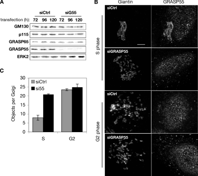
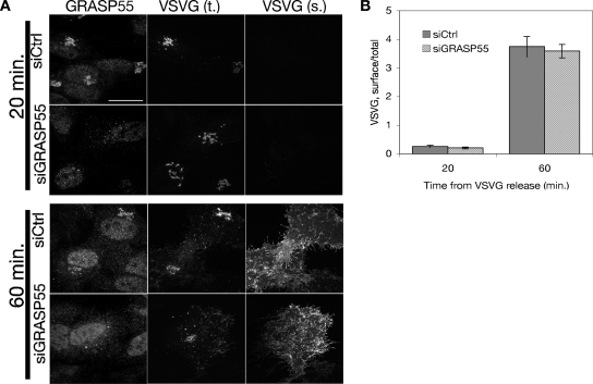
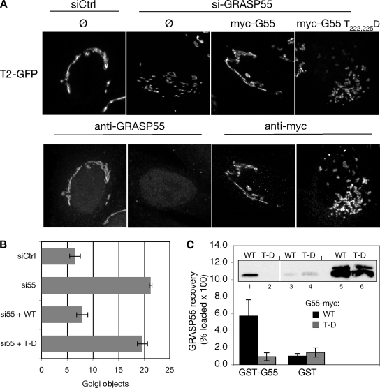
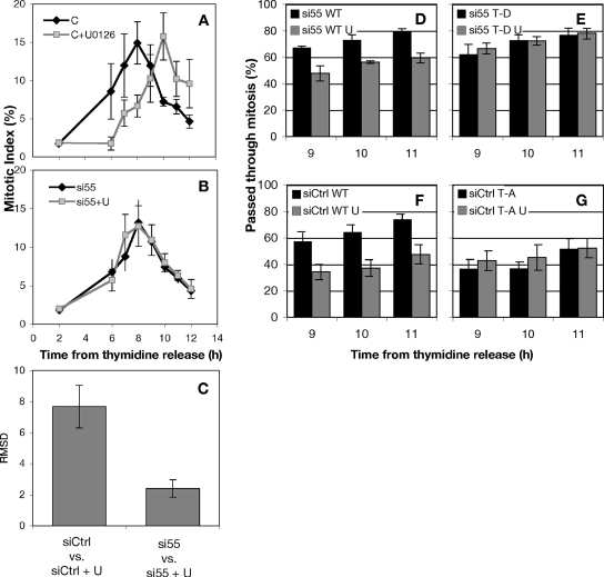
Recent work indicates that mitogen-activated protein kinase kinase (MEK)1 signaling at the G2/M cell cycle transition unlinks the contiguous mammalian Golgi apparatus and that this regulates cell cycle progression. Here, we sought to determine the role in this pathway of Golgi reassembly protein (GRASP)55, a Golgi-localized target of MEK/extracellular signal-regulated kinase (ERK) phosphorylation at mitosis. In support of the hypothesis that GRASP55 is inhibited in late G2 phase, causing unlinking of the Golgi ribbon, we found that HeLa cells depleted of GRASP55 show a fragmented Golgi similar to control cells arrested in G2 phase. In the absence of GRASP55, Golgi stack length is shortened but Golgi stacking, compartmentalization, and transport seem normal. Absence of GRASP55 was also sufficient to suppress the requirement for MEK1 in the G2/M transition, a requirement that we previously found depends on an intact Golgi ribbon. Furthermore, mimicking mitotic phosphorylation of GRASP55 by using aspartic acid substitutions is sufficient to unlink the Golgi apparatus in a gene replacement assay. Our results implicate MEK1/ERK regulation of GRASP55-mediated Golgi linking as a control point in cell cycle progression.


Figures

Figure 1.
Figure 1.
Depletion of GRASP55 mimics G2 phase Golgi fragmentation. (A) Extracts from HeLa cells lysed 72, 96, and 120 h after transfection with either control or GRASP55 siRNA were analyzed by immunoblotting using anti-GRASP55 or antibodies against the indicated proteins. (B) Control- or GRASP55 siRNA-transfected cells were synchronized in S phase using thymidine or at the G2/M transition point by using olomoucine II, and the Golgi was visualized by staining for giantin (left) or GRASP55 (right). Bar, 4 μm. (C) Distinct Golgi objects per cell were estimated using fixed acquisition parameters and image thresholding, followed by counting using the Analyze Objects function in ImageJ (n = 4, ±SEM).
Figure 2.
Figure 2.
GRASP55 depletion severs the connections between Golgi stacks and inhibits normal protein glycosylation. (A) Representative cells stably expressing GalNAcT2-GFP were tested for Golgi integrity by photobleaching a Golgi segment with a brief laser pulse and measuring the return of Golgi fluorescence to the bleached spot. Bar, 10 μm. Corresponding movies are provided (see Supplemental Figures S1 and S2). (B) Fluorescence recovery was quantified by dividing GFP fluorescence within the bleached spot by the fluorescence within a nearby unbleached region in the same Golgi object, after subtracting background fluorescence from both. Each curve was further normalized by bracketing values between a minimum of 0 and a maximum of 1 (n = 3, ≥12 cells each, ±SEM). (C) To test the glycolytic processing of Golgi cargo, live, nonpermeabilized, control- or GRASP55-depleted cells were stained with fluorescently tagged GSII lectin and then rinsed, fixed and processed for immunofluorescence by using GP73 as a Golgi marker. Bar, 4 μm.
Figure 3.
Figure 3.
GRASP55 is required for the reformation of Golgi ribbons. (A and B) HeLa cells treated with nocodazole for 3 h to redistribute the Golgi to peripheral ministacks were rinsed repeatedly, and then they were transferred to chambers for live imaging. Confocal image stacks were collected every 30 s and collapsed into 2D images for analysis (larger frames). Smaller frames represent enlargements shown at 60-s intervals to illustrate tubulation and membrane capture that was frequently observed during Golgi reassembly. Movies are representative of at least three independent experiments per treatment. Corresponding movies are provided (Supplemental Figures S3 and S4).
Figure 4.
Figure 4.
GRASP55 selectively labels tubules that connect reassembling Golgi elements. To determine the relative abundance of golgins and resident proteins on cisternal bridging tubules, HeLa cells expressing GRASP55-GFP were fixed at designated time points after nocodazole washout, and then they were processed for immunofluorescence by using and antibody against the cis-Golgi resident protein GP73. Arrows, tubules labeled with either GRASP55 or GP73. Results are representative of three separate experiments. A movie of GRASP55-GFP labeled Golgi assembly after nocodazole washout is provided as a supplemental movie (see Supplemental Figure S5).
Figure 5.
Figure 5.
Transmission electron microscopy of GRASP55-depleted Golgi. (A and B) Representative TEM thin sections showing Golgi from control and GRASP55-depleted cells. Bar, 500 nm. (C) Quantitative representation of Golgi ribbon length in thin sections from control and GRASP55-depleted cells. The length of membrane profiles was estimated using the freehand line tool in ImageJ (n = 3, ≥20 profiles each, ±SEM). (D) Golgi stack lengths from a representative experiment were divided into size class bins and presented in histogram format to illustrate the distinct distributions of stack lengths in the presence (black diamonds) and absence (gray squares) of GRASP55. Note that despite the variation evident in the histogram, the error in panel C is small because the average stack length was consistent across multiple experiments. (E) Masks were drawn over Golgi membranes taken from a contiguous stack of TEM serial sections (representative sections are shown in insets) and reassembled into a 3D volume. The full 3D representations are provided as supplemental movies (see Supplemental Figures S6 and S7).
Figure 6.
Figure 6.
Evidence of marker compartmentalization after GRASP55 depletion. (A) HeLa cells expressing the medial resident protein GalNAcT2-GFP and either control (top) or GRASP55 depleted (middle) or treated with nocodazole for 3 h (bottom) were fixed and processed for immunofluorescence by using antibodies against the cis-Golgi marker GPP130. Bar, 4 μm. (B) An enlarged selection from each of the RGB images above (white squares) is presented to emphasize the physically distinct cis and medial staining in all three treatments. (C) Representative linear transects through Golgi (white lines in A) showing the distinct fluorescence intensity profiles for GPP130 (black traces) and GalNAcT2-GFP (gray traces).
Figure 7.
Figure 7.
Anterograde traffic through the Golgi in GRASP55-depleted cells. (A) Golgi transport was measured after release of ts045 VSVG-GFP from a 40°C ER block by shift to 32°C. Total VSVG was measured using GFP fluorescence, whereas surface VSVG was measured by staining live cells with a monoclonal antibody against a lumenal fragment of VSVG. (B) Surface and total fluorescence values were quantified and represented as ratios to indicate the relative amount of VSVG on the plasma membrane. Data shown are representative values from one of three experiments.
Figure 8.
Figure 8.
Aspartic acid substitution at threonines 222 and 225 inhibits Golgi linking and oligomerization activities of GRASP55. (A) Golgi ribbon integrity was assessed in control (siCtrl) or GRASP55 (siGRASP55) knockdown cells without gene replacement (Ø) or after replacement with myc-tagged GRASP55 or GRASP55 T222,225D. Images show GalNAcT2-GFP fluorescence (top) and in the same cells either anti-GRASP55 staining or anti-myc staining (bottom). Bar, 4 μm. (B) The number of Golgi objects per cell was counted using the automated protocol described in the Figure 1 legend (n = 4, ±SEM). (C) Extracts from HeLa cells transfected with either wild-type GRASP55 (WT) or GRASP55 T222,225D (T-D) were incubated with GST-GRASP55 (lanes 1 and 2) or GST alone (lanes 3 and 4) immobilized on glutathione-agarose beads. Recovery was determined by immunoblotting with anti-myc antibodies. Values shown represent the percentage of loaded protein (lanes 5–6) bound to beads × 100 (n = 5, ±SEM).
Figure 9.
Figure 9.
MEK1 promotes mitosis at G2/M by phosphorylation of GRASP55 at T222 and T225. (A) The percentage of cells in mitosis (mitotic index) was measured after thymidine release. The MEK1/2 inhibitor U0126 or 0.05% dimethyl sulfoxide carrier were added at 6 h after release. Tandem experiments were carried out in cells transfected with control siRNA (A and B) or siRNA against GRASP55 (B). (C) The degree to which MEK1 inhibition delays mitosis was quantified using an RMSD comparison of control and U0126-treated cells. (D–G) A postmitotic couplet assay was used to estimate the abundance of postmitotic HeLa cells expressing wild-type (D) or nonphosphorylatable GRASP55 (G55 T222,225A; E). Conversely, a functional gene replacement strategy was used (Puthenveedu et al., 2006) to compare wild-type expressors (F) with cells expressing a mutant GRASP55 designed to mimic mitotic ERK phosphorylation (G55 T222,225D; G).

Similar articles

Cited by

References

    1. Allan V. J., Thompson H. M., McNiven M. A. Motoring around the Golgi. Nat. Cell Biol. 2002;4:E236–E242. - PubMed
    1. Angata K., Lee W., Mitoma J., Marth J. D., Fukuda M. Cellular and molecular analysis of neural development of glycosyltransferase gene knockout mice. Methods Enzymol. 2006;417:25–37. - PubMed
    1. Bachert C., Fimmel C., Linstedt A. D. Endosomal trafficking and proprotein convertase cleavage of cis Golgi protein GP73 produces marker for hepatocellular carcinoma. Traffic. 2007;8:1415–1423. - PubMed
    1. Barr F. A., Preisinger C., Kopajtich R., Korner R. Golgi matrix proteins interact with p24 cargo receptors and aid their efficient retention in the Golgi apparatus. J. Cell Biol. 2001;155:885–891. - PMC - PubMed
    1. Barr F. A., Puype M., Vandekerckhove J., Warren G. GRASP65, a protein involved in the stacking of Golgi cisternae. Cell. 1997;91:253–262. - PubMed

Publication types

LinkOut - more resources