. 2008 Jul;295(1):C288-92.
doi: 10.1152/ajpcell.00104.2008. Epub 2008 Apr 23.

Functional interaction of regulatory factors with the Pgc-1alpha promoter in response to exercise by in vivo imaging

Affiliations

Functional interaction of regulatory factors with the Pgc-1alpha promoter in response to exercise by in vivo imaging

Takayuki Akimoto et al. Am J Physiol Cell Physiol. 2008 Jul.

Abstract

Real-time optical bioluminescence imaging is a powerful tool for studies of gene regulation in living animals. To elucidate exercise-induced signaling/transcriptional control of the peroxisome proliferator-activated receptor-gamma coactivator-1alpha (Pgc-1alpha) gene in skeletal muscle, we combined this technology with electric pulse-mediated gene transfer to cotransfect the Pgc-1alpha reporter gene with plasmid DNA encoding mutant/deletion forms of putative regulatory factors and, thereby, assess the responsiveness of the promoter to skeletal muscle contraction. We show that each of the myocyte enhancer factor 2 sites on the Pgc-1alpha promoter is required for contractile activity-induced Pgc-1alpha transcription. The responsiveness of the Pgc-1alpha promoter to contractile activity could be completely blocked by overexpression of the dominant-negative form of activating transcription factor 2 (ATF2), the signaling-resistant form of histone deacetylase (HDAC) 5 (HDAC5), or protein kinase D (PKD), but not by HDAC4. These findings provide in vivo evidence for functional interactions between PKD/HDAC5 and ATF2 regulatory factors and the Pgc-1alpha gene in adult skeletal muscle.


Figures
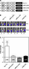
Fig. 1.
Fig. 1.
Peroxisome proliferator-activated receptor-γ coactivator-1α (Pgc-1α) promoter activity is dependent on each of the myocytic enhancer factor 2 (MEF2) and cAMP response element (CRE) sites. After transfection of both tibialis anterior (TA) muscles by Pgc-1αL or its derivative mutant constructs, the motor nerve of one of the TA muscles was stimulated for bioluminescence imaging analysis. A: schematic presentation of plasmid DNAs. Pgc-1α promoter (3.1 kb) is presented as a solid line with two MEF2 sites and one CRE site. Crosses denote site-directed mutagenesis as described elsewhere (6, 12). B: in vivo imaging of luciferase activity in mouse TA muscles from Pgc-1αL and its mutant derivatives ΔMEF2(−2901), ΔMEF2(−1539), and ΔCRE(−222). Imaging analysis was performed 10 days after gene transfer. Pseudocolors overlaid on the image indicate intensity of luminescent signals from luciferase reporter gene activity. Animals' right TA muscles were stimulated (S), and their left TA muscles were sham-operated without stimulation and used as reference control (C). C: quantification of luciferase activity in TA muscles. Dashed horizontal line denotes basal level before stimulation. Values are means ± SE; n = 6–8. **P < 0.01 vs. poststimulation values.
Fig. 2.
Fig. 2.
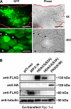
Efficient gene expression following electric pulse-mediated gene transfer. Plasmid DNA [65 μg of pEGFP-N1 or 15 μg of Pgc-1αL and 50 μg of plasmid DNA encoding an empty control vector (pCI-neo) or a mutant/deletion form of regulatory factor] was transfected into TA muscles, which were visualized by epifluorescent microscopy (A) and subjected to homogenization and immunobloting analysis (B). A: TA muscle transfected with pEGFP-N1 was harvested and sectioned for phase contrast (Phase) and fluorescent microscopic examination for expression of green fluorescent protein (GFP). There were ∼60% GFP-positive myofibers. Red lines outline boundary of muscle sections. *, Myofibers that are negative for GFP signals. Scale bars, 250 and 50 μm for ×4 and ×40 images, respectively. B: expression of ATF2ΔN, HDAC5(S/A), HDAC4(S/A), and PKD(K/W) could be easily detected in transfected TA muscles by immunoblot using anti-FLAG or anti-hemagglutinin (HA) antibodies for tagged proteins and anti-α-tubulin antibodies for protein loading. ATF2, activating transcription factor 2; HDAC, histone deacetylase; PKD, protein kinase D.
Fig. 3.
Fig. 3.
Contractile activity-dependent functional interaction between Pgc-1α promoter and upstream regulatory factors. A: in vivo imaging of luciferase activity in mouse TA muscles from Pgc-1αL cotransfected with an empty control vector (pCI-neo), ATF2ΔN, HDAC5(S/A), HDAC4(S/A), or PKD(K/W) before (Pre) and 2 h after (Post) low-frequency (10-Hz) nerve stimulation (2 h). Animals' right TA muscles were stimulated (S), and their left TA muscles were sham-operated without stimulation and used as reference control (C). B: quantitative analysis of luciferase activity from Pgc-1αL. Dashed horizontal line denotes basal level before stimulation. Values are means ± SE. *P < 0.05; **P < 0.01 vs. poststimulation values.
Fig. 4.
Fig. 4.
Model for exercise-induced transcription of the Pgc-1α gene in skeletal muscle. Increased neuromuscular activities elicit signals leading to activation of the p38 MAPK pathway and transcriptional upregulation of the Pgc-1α gene through ATF2 (1, 34) and MEF2 (11, 37). Activation of the p38 MAPK pathway can also promote Pgc-1α transcription by inhibiting p160 and derepressing PGC-1α protein function (8), exerting a positive regulation of Pgc-1α gene transcription through MEF2 (12, 24). In parallel, exercise also promotes Pgc-1α transcription through PKD/HDAC5-mediated derepression of MEF2 function (6). These signaling-transcription coupling cascades integrate contractile and metabolic cues from neuromuscular activity to control expression of the Pgc-1α gene in skeletal muscle adaptation. Regulatory factors/cis elements, the functional role of which has been tested and confirmed in the present study, are shown as gray boxes with solid lines. Other relevant regulatory factors that were not investigated in the present study are shown as open boxes with dashed lines.

References

    1. Akimoto T, Pohnert SC, Li P, Zhang M, Gumbs C, Rosenberg PB, Williams RS, Yan Z. Exercise stimulates Pgc-1α transcription in skeletal muscle through activation of the p38 MAPK pathway. J Biol Chem 280: 19587–19593, 2005. - PubMed
    1. Akimoto T, Sorg BS, Yan Z. Real-time imaging of peroxisome proliferator-activated receptor-γ coactivator-1α promoter activity in skeletal muscles of living mice. Am J Physiol Cell Physiol 287: C790–C796, 2004. - PubMed
    1. Baar K, Wende AR, Jones TE, Marison M, Nolte LA, Chen M, Kelly DP, Holloszy JO. Adaptations of skeletal muscle to exercise: rapid increase in the transcriptional coactivator PGC-1. FASEB J 16: 1879–1886, 2002. - PubMed
    1. Boppart MD, Asp S, Wojtaszewski JF, Fielding RA, Mohr T, Goodyear LJ. Marathon running transiently increases c-Jun NH2-terminal kinase and p38 activities in human skeletal muscle. J Physiol 526: 663–669, 2000. - PMC - PubMed
    1. Boppart MD, Hirshman MF, Sakamoto K, Fielding RA, Goodyear LJ. Static stretch increases c-Jun NH2-terminal kinase activity and p38 phosphorylation in rat skeletal muscle. Am J Physiol Cell Physiol 280: C352–C358, 2001. - PubMed

Publication types

MeSH terms