. 2008 Jul;23(7):1118-28.
doi: 10.1359/jbmr.080304.

Age-related changes in the osteogenic differentiation potential of mouse bone marrow stromal cells

Affiliations

Age-related changes in the osteogenic differentiation potential of mouse bone marrow stromal cells

Weixi Zhang et al. J Bone Miner Res. 2008 Jul.

Abstract

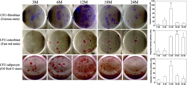
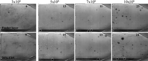
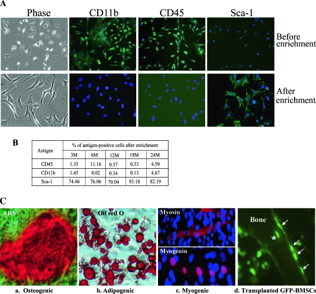
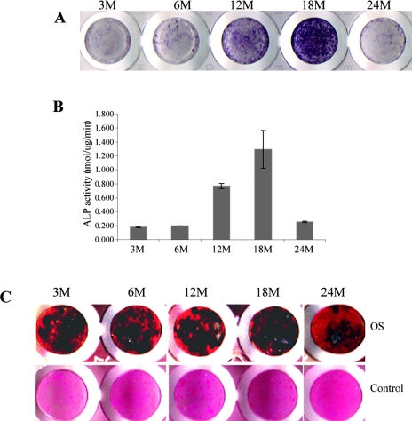
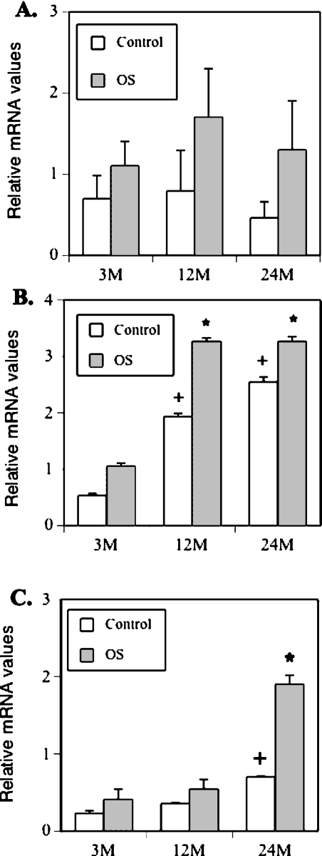
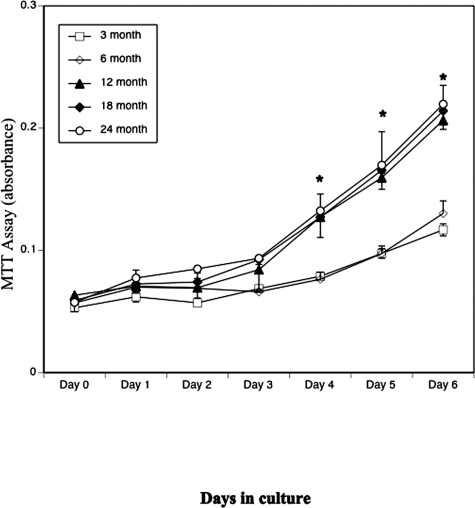
Age-dependent bone loss has been well documented in both human and animal models. Although the underlying causal mechanisms are probably multifactorial, it has been hypothesized that alterations in progenitor cell number or function are important. Little is known regarding the properties of bone marrow stromal cells (BMSCs) or bone progenitor cells during the aging process, so the question of whether aging alters BMSC/progenitor osteogenic differentiation remains unanswered. In this study, we examined age-dependent changes in bone marrow progenitor cell number and differentiation potential between mature (3 and 6 mo old), middle-aged (12 and 18 mo old), and aged (24 mo old) C57BL/6 mice. BMSCs or progenitors were isolated from five age groups of C57BL/6 mice using negative immunodepletion and positive immunoselection approaches. The osteogenic differentiation potential of multipotent BMSCs was determined using standard osteogenic differentiation procedures. Our results show that both BMSC/progenitor number and differentiation potential increase between the ages of 3 and 18 mo and decrease rapidly thereafter with advancing age. These results are consistent with the changes of the mRNA levels of osteoblast lineage-associated genes. Our data suggest that the decline in BMSC number and osteogenic differentiation capacity are important factors contributing to age-related bone loss.


Figures

FIG. 1
FIG. 1
Assays for the number of colony-forming units for fibroblasts, osteoblasts, and adipocytes. Bone marrow cells from indicated ages of mice were cultured for 14 days in MesenCult media and stained with Giemsa (CFU-fibroblast), cultured for 12 days in osteogenic induction media and stained with Fast Red Violet LB (CFU-osteoblast), or cultured in adipogenic induction media for 2 days followed by a 9-day incubation in maintenance media and then stained with Oil Red O (CFU-adipocyte). Quantitative results for these assays are shown as bar graphs on right. CFU-fibroblast and CFU-osteoblast assays were performed in duplicate in 6-well plates with 5 × 10⁵ nucleated cells/well. CFU-adipocyte assays were performed in triplicate in 24-well plates with 1 × 10⁶ nucleated cells/well. Only one representative well is shown for each age group. These experiments were repeated in triplicate in two separate experiments (*p < 0.001 compared with 3 mo).
FIG. 2
FIG. 2
Effect of guinea pig feeder cells on CFU-fibroblast colony-forming efficiency; 3 × 10⁶ to 10 × 106 nucleated mouse bone marrow cells were seeded in two sets of 25-cm2 plastic culture flasks. Top panels (Feeder layer): 3 h after seeding, unattached cells from one set of the flasks were removed by aspiration, washed vigorously three times with DMEM. 1 × 10⁷ nucleated guinea pig bone marrow cells, freshly prepared and immediately γ-irradiated (6000 R), were added to each flask (5-ml volume). The cultures were incubated for 10 days and stained with Giemsa as in Fig. 1. Bottom panels (20% FBS): 24 h after seeding, the media in the other set of the flasks were removed, and the flasks were washed gently two times with DMEM. Five milliliters of DMEM supplemented with 20% FBS was added to each flask, incubated, and stained as above. Colonies (>50 cells in size) were counted visually, and the number of colonies in each flask is indicated. The white circles are used to highlight individual colonies.
FIG. 3
FIG. 3
Characterization of BMSCs. (A) Marrow cell cultures (top) and antibody-enriched BMSCs from 18-mo-old mice (bottom) were immuno-labeled with FITC-conjugated monoclonal antibodies as indicated and detected using a fluorescence microscope. (B) FACS analysis of enriched BMSCs. BMSCs from indicated ages of mice were labeled with FITC-conjugated monoclonal antibodies (CD45, CD11b, and Sca-1, respectively) and analyzed. Percentages of cells positive for CD45, CD11b, and Sca-1 are shown. This experiment was performed in triplicate. (C) Multipotentiality of enriched BMSCs. BMSCs isolated from 18-mo-old mice were exposed to osteogenic, adipogenic, and myogenic differentiation media. (a) Cells were treated with osteogenic supplements for 21 days and stained with ARS for mineralized bone nodules. (b) Cells were treated with adipogenic induction media for 2 days, cultured in maintenance media for 14 days, and stained with Oil Red O. (c) Cells were treated with 5-azacytidine (5-Aza) for 24 h, cultured in regular growth media for 21 days, and immuno-labeled with indicated antibodies. These experiments were performed a minimum of three times in triplicate. (d) Bone tissue labeled with GFP antibody to show osteoblast-like lining cells (arrows) 6 wk after injection of GFP-BMSC into mouse tibias. Star indicates GFP-BMSC embedded in the bone. These experiments were repeated at least three times with similar results.
FIG. 4
FIG. 4
Effect of aging on BMSC osteogenic differentiation. BMSCs from indicated ages of mice were cultured in osteogenic induction media for 10 or 21 days and assayed for ALP activity or mineralization. (A and B) After 10 days of treatment, cells were fixed with 3.7% formaldehyde, and stained with SIGMA FAST BCIP/NBT Buffered Substrate (A), or lysed in 0.05% Triton X-100 directly without fixing and assayed for ALP activity (B). ALP activity is given in Sigma units normalized to protein content, where 1 Sigma unit is equivalent to the enzyme activity needed to release 1 mol of p-nitrophenol per hour. The protein concentration was determined using Bradford reagent (*p < 0.01; + p < 0.001 compared with 3 mo). The experiment was done in triplicate in two separate experiments. (C) After 21 days of treatment, cells were fixed with formaldehyde and stained with silver nitrate for mineralized nodules (von Kossa staining). The cells were counterstained with eosin. These experiments were performed a minimum of three times in triplicate. Cells cultured in MDEM did not mineralize. Only one representative stained wells for each age group is shown.
FIG. 5
FIG. 5
Real-time RT-PCR analysis of osteoblast-specific gene expression. BMSCs from 3-, 12-, and 24-mo-old mice were treated with or without osteogenic induction media for 14 days and harvested for total RNA isolation. The mRNA levels of Runx2/Cbfa1, type I collagen (Col), and osteocalcin were analyzed by real-time RT-PCR. The PCR reactions were performed in triplicate for each sample in two separate experiments (+ p < 0.001; *p < 0.01 compared with 3 mo).
FIG. 6
FIG. 6
Effect of aging on BMSC proliferation. BMSCs from 3-, 6-, 12-, 18-, and 24-mo-old mice were plated in triplicate in 96-well plates at a density of 1 × 10⁴ cells/cm2, and cell proliferation was assayed using a Cell Growth Determination kit for 7 consecutive days. These experiments were repeated in triplicate in two separate experiments (*p < 0.01 compared with 3 mo).

References

    1. Ferguson VL, Ayers RA, Bateman TA, Simske SJ. Bone development and age-related bone loss in male C57BL/6J mice. Bone. 2003;33:387–398. - PubMed
    1. Gudmundsdottir SL, Indridason OS, Franzson L, Sigurdsson G. Age-related decline in bone mass measured by dual-energy X-ray absorptiometry and quantitative ultrasound in a population-based sample of both sexes: Identification of useful ultrasound thresholds for osteoporosis screening. J Clin Densitom. 2005;8:80–86. - PubMed
    1. Hamrick MW, Shi X, Zhang W, Pennington C, Thakore H, Haque M, Kang B, Isales CM, Fulzele S, Wenger KH. Loss of myostatin (GDF8) function increases osteogenic differentiation of bone marrow-derived mesenchymal stem cells but the osteogenic effect is ablated with unloading. Bone. 2007;40:1544–1553. - PMC - PubMed
    1. Rupprecht M, Pogoda P, Mumme M, Rueger JM, Puschel K, Amling M. Bone microarchitecture of the calcaneus and its changes in aging: A histomorphometric analysis of 60 human specimens. J Orthop Res. 2006;24:664–674. - PubMed
    1. Manolagas SC. Birth and death of bone cells: Basic regulatory mechanisms and implications for the pathogenesis and treatment of osteoporosis. Endocr Rev. 2000;21:115–137. - PubMed

Publication types