

Establishing humanized mice using stem cells: maximizing the potential
- PMID: 18435804
- PMCID: PMC2453213
- DOI: 10.1111/j.1365-2249.2008.03659.x
Establishing humanized mice using stem cells: maximizing the potential
Abstract
Studies on physiology and pathology as they relate to the immune system draw heavily upon rodent models. With the increasing impetus provided by initiatives in translational medicine, the demand for ever more sophisticated, 'humanized' murine models is greater than ever. However, the design and implementation of studies in such mice is far from trivial. Here we provide a technical perspective on the increasing interest in developing humanized mice. We give examples of primary data starting with the routine procurement of human donor material, through CD34(+) cell purification prior to engraftment to injection into immunocompromised mice. Our goal is to provide practical advice to the many investigators who may be commencing or considering such studies.
Figures
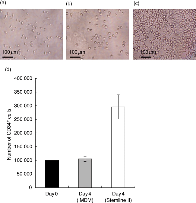
 ) and day 4 using Stemline II (□).
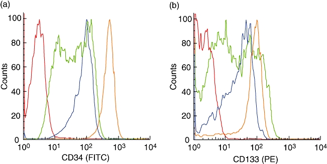
 ), CD34+ after 4 days in IMDM medium (
 ) and CD34+ after 4 days in Stemline II medium (
 ). Isotype control (
 ). (b) Comparison of CD133 expression between fresh CD34+ cells (
 ), CD34+ after 4 days in IMDM medium (
 ) and CD34+ after 4 days in Stemline II medium (
 ). Isotype control (
 ). FITC, fluorescein isothiocyanate; PE, phycoerythrin.
References
-
- Serreze DV, Chen YG. Of mice and men: use of animal models to identify possible interventions for the prevention of autoimmune type 1 diabetes in humans. Trends Immunol. 2005;26:603–7. - PubMed
-
- Roep BO, Atkinson M. Animal models have little to teach us about type 1 diabetes: 1. In support of this proposal. Diabetologia. 2004;47:1650–6. - PubMed
-
- Roep BO, Atkinson M, von Herrath M. Satisfaction (not) guaranteed: re-evaluating the use of animal models of type 1 diabetes. Nat Rev Immunol. 2004;4:989–97. - PubMed
-
- Mosier DE, Gulizia RJ, Baird SM, Wilson DB. Transfer of a functional human immune system to mice with severe combined immunodeficiency. Nature. 1988;335:256–9. - PubMed
-
- Bosma GC, Custer RP, Bosma MJ. A severe combined immunodeficiency mutation in the mouse. Nature. 1983;301:527–30. - PubMed
Publication types
MeSH terms
Substances
Grants and funding
LinkOut - more resources
Full Text Sources
Other Literature Sources
