. 2008 Apr 25:3:17.
doi: 10.1186/1749-799X-3-17.

Fibrin and poly(lactic-co-glycolic acid) hybrid scaffold promotes early chondrogenesis of articular chondrocytes: an in vitro study

Affiliations

Fibrin and poly(lactic-co-glycolic acid) hybrid scaffold promotes early chondrogenesis of articular chondrocytes: an in vitro study

Munirah Sha'ban et al. J Orthop Surg Res. .

Abstract

Background: Synthetic- and naturally derived- biodegradable polymers have been widely used to construct scaffolds for cartilage tissue engineering. Poly(lactic-co-glycolic acid) (PLGA) are bioresorbable and biocompatible, rendering them as a promising tool for clinical application. To minimize cells lost during the seeding procedure, we used the natural polymer fibrin to immobilize cells and to provide homogenous cells distribution in PLGA scaffolds. We evaluated in vitro chondrogenesis of rabbit articular chondrocytes in PLGA scaffolds using fibrin as cell transplantation matrix.

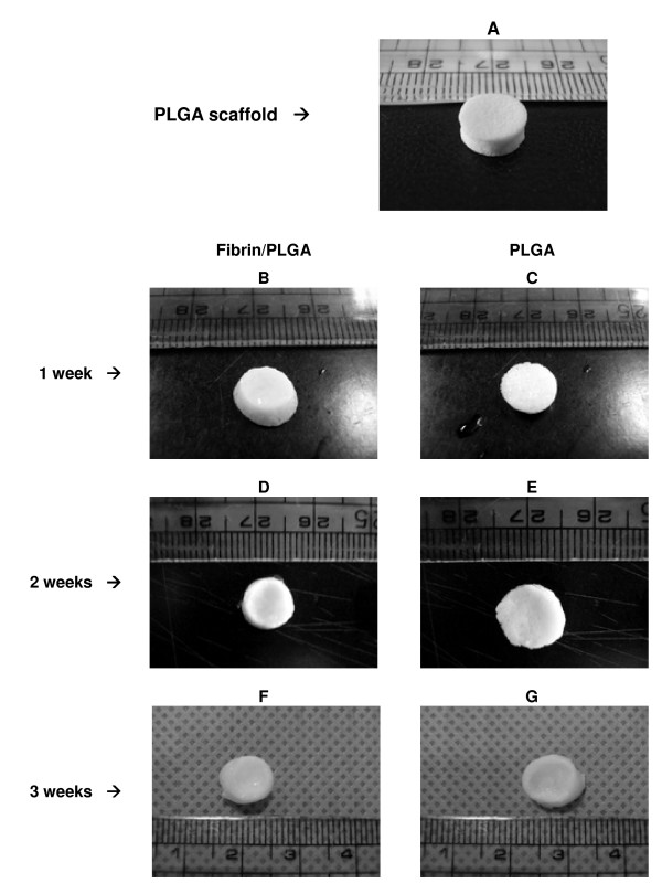
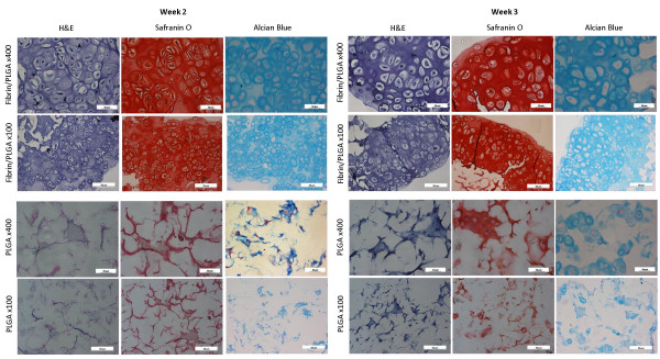
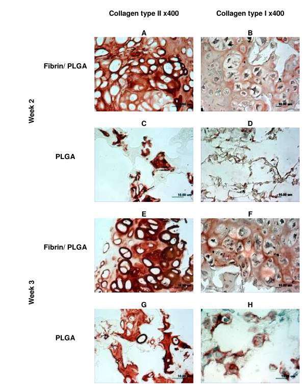
Methods: PLGA scaffolds were soaked in chondrocytes-fibrin suspension (1 x 10(6) cells/scaffold) and polymerized by dropping thrombin-calcium chloride (CaCl2) solution. PLGA-seeded chondrocytes was used as control. All constructs were cultured for a maximum of 21 days. Cell proliferation activity was measured at 1, 3, 7, 14 and 21 days in vitro using 3-(4,5-dimethylthiazole-2-yl)-2-, 5-diphenyltetrazolium-bromide (MTT) assay. Morphological observation, histology, immunohistochemistry (IHC), gene expression and sulphated-glycosaminoglycan (sGAG) analyses were performed at each time point of 1, 2 and 3 weeks to elucidate in vitro cartilage development and deposition of cartilage-specific extracellular matrix (ECM).

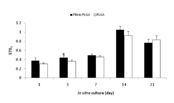
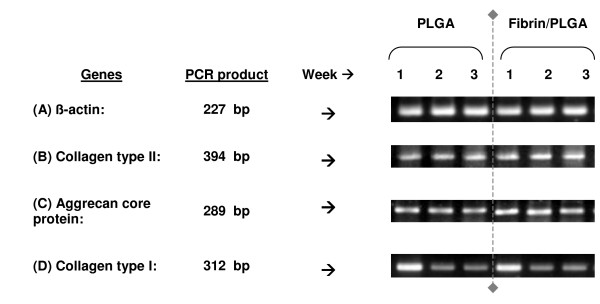
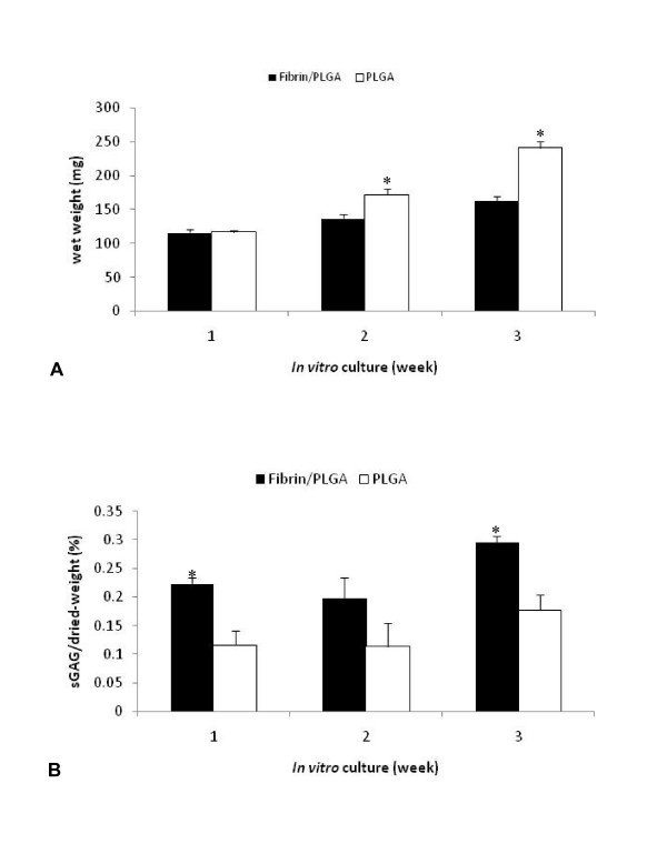
Results: Cell proliferation activity was gradually increased from day-1 until day-14 and declined by day-21. A significant cartilaginous tissue formation was detected as early as 2-week in fibrin/PLGA hybrid construct as confirmed by the presence of cartilage-isolated cells and lacunae embedded within basophilic ECM. Cartilage formation was remarkably evidenced after 3 weeks. Presence of cartilage-specific proteoglycan and glycosaminoglycan (GAG) in fibrin/PLGA hybrid constructs were confirmed by positive Safranin O and Alcian Blue staining. Collagen type II exhibited intense immunopositivity at the pericellular matrix. Chondrogenic properties were further demonstrated by the expression of genes encoded for cartilage-specific markers, collagen type II and aggrecan core protein. Interestingly, suppression of cartilage dedifferentiation marker; collagen type I was observed after 2 and 3 weeks of in vitro culture. The sulphated-glycosaminoglycan (sGAG) production in fibrin/PLGA was significantly higher than in PLGA.

Conclusion: Fibrin/PLGA promotes early in vitro chondrogenesis of rabbit articular chondrocytes. This study suggests that fibrin/PLGA may serve as a potential cell delivery vehicle and a structural basis for in vitro tissue-engineered articular cartilage.


Figures

Figure 1
Figure 1
Measurement of cell proliferation activity of in vitro constructs. Fibrin/PLGA and PLGA construct exhibited similar growth pattern in vitro. Cells proliferation was gradually increased until day-14. Fibrin/PLGA showed a significant higher (p < 0.05) cells proliferation than PLGA at day-3 (*). Cells proliferation activity had declined by day-21.
Figure 2
Figure 2
Macroscopic observation of in vitro constructs. Figure 2A represents PLGA scaffold which was designed in the shape of cylindrical disc. Fibrin/PLGA constructs (Figure 2B) and PLGA construct (Figure 2C) was morphologically similar after 7 days in culture. Fibrin/PLGA construct (Figure 2D) showed slightly smooth and glistening morphology when compared to PLGA (Figure 2E) after 14 days. By week 3, fibrin/PLGA construct appeared whiter, smoother and glistening (Figure 2F) than PLGA (Figure 2G).
Figure 3
Figure 3
Histological evaluation of in vitro constructs. Fibrin/PLGA constructs showed superior histological features of cartilage-like tissue compared to PLGA. Differences between fibrin/PLGA (Figure 3A, B, C and Figure 3G, H, I) and PLGA (Figure 3D, E, F and Figure 3J, K, L) were clearly visible in term of overall cartilaginous tissue formation, cells organization and ECM distribution. The fibrin/PLGA constructs was intensely stained with Safranin O for accumulated proteoglycan and Alcian Blue for GAG at 2 weeks and greatest at 3 weeks.
Figure 4
Figure 4
Immunohistochemistry analysis of in vitro constructs. As shown in Figure 4A, fibrin/PLGA exhibited strong immunopositivity of collagen type II which mainly localized at the pericellular and inter-territorial matrix. Minimal collagen type II expression could be observed in the PLGA construct (Figure 4C). After 3 weeks, collagen type II expression was maintained in fibrin/PLGA (Figure 4E) and PLGA (Figure G). Collagen type I in fibrin/PLGA constructs showed moderate immunopositivity at week-2 (Figure 4B) and week-3 (Figure 4F), as did PLGA (Figure 4D, Figure 4H).
Figure 5
Figure 5
Cartilage-specific phenotypic expression analysis. The expression of genes encoded the cartilage-specific markers; collagen type II and aggrecan core protein was steadily expressed in fibrin/PLGA and PLGA. Interestingly, suppression of collagen type I was observed in fibrin/PLGA and PLGA at 2 weeks and 3 weeks. β-actin gene was steadily expressed in all samples to verify the analysis was reliable and successful.
Figure 6
Figure 6
Sulphated-glycosaminoglycan (sGAG) production assay. The wet weight (Figure 6A) and sGAG production (Figure 6B) of the in vitro constructs were measured at 1, 2, and 3 weeks of culture, respectively. After 2 and 3 weeks in vitro, PLGA demonstrated significantly higher wet weight (p < 0.05) compared to fibrin/PLGA. The sGAG production in fibrin/PLGA construct was superior to PLGA. Relative sGAG contents (%) were significantly higher (p < 0.05) in fibrin/PLGA than PLGA at 1 week and 3 weeks.

Similar articles

Cited by

References

    1. Brittberg M, Lindahl A, Nilsson A, Ohlsson C, Isaksson O, Peterson L. Treatment of deep cartilage defects in the knee with autologous chondrocyte transplantation. N Engl J Med. 1994;331:889–95. doi: 10.1056/NEJM199410063311401. - DOI - PubMed
    1. Shao XX, Hutmacher DW, Ho ST, Goh JCH, Lee EH. Evaluation of a hybrid scaffold/cell construct repair of high load bearing osteochondral defects in rabbits. Biomaterials. 2006;27:1071–80. doi: 10.1016/j.biomaterials.2005.07.040. - DOI - PubMed
    1. Willers C, Chen J, Wood D, Xu J, Zheng MH. Autologous chondrocytes implantation with collagen bioscaffold for the treatment of osteochondral defects in rabbits. Tissue Eng. 2005;11:1065–76. doi: 10.1089/ten.2005.11.1065. - DOI - PubMed
    1. Ito Y, Ochi M, Adachi N, Sugawara K, Yanada S, Ikada Y, Ronakorn P. Repair of osteochondral defect with tissue engineered chondral plug in rabbit model. Arthroscopy. 2005;21:1155–63. - PubMed
    1. Niederauer GG, Silva MA, Leatherbury NC, Korvick DL, Harroff HH, Ehler WC, Dunn CJ, Kiewsetter K. Evaluation of multiphase implants for repair of focal osteochondral defects in goats. Biomaterials. 2000;21:2561–74. doi: 10.1016/S0142-9612(00)00124-1. - DOI - PubMed

LinkOut - more resources