. 2008 Sep;87(8-9):721-34.
doi: 10.1016/j.ejcb.2008.02.011. Epub 2008 Apr 23.

The Rsu-1-PINCH1-ILK complex is regulated by Ras activation in tumor cells

Affiliations

The Rsu-1-PINCH1-ILK complex is regulated by Ras activation in tumor cells

Gerard W Dougherty et al. Eur J Cell Biol. 2008 Sep.

Abstract

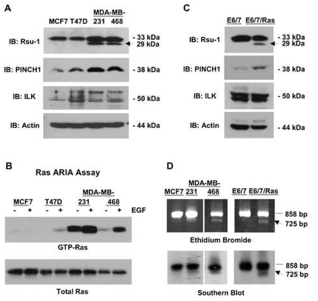
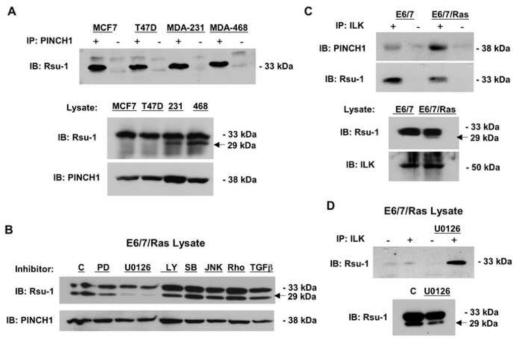
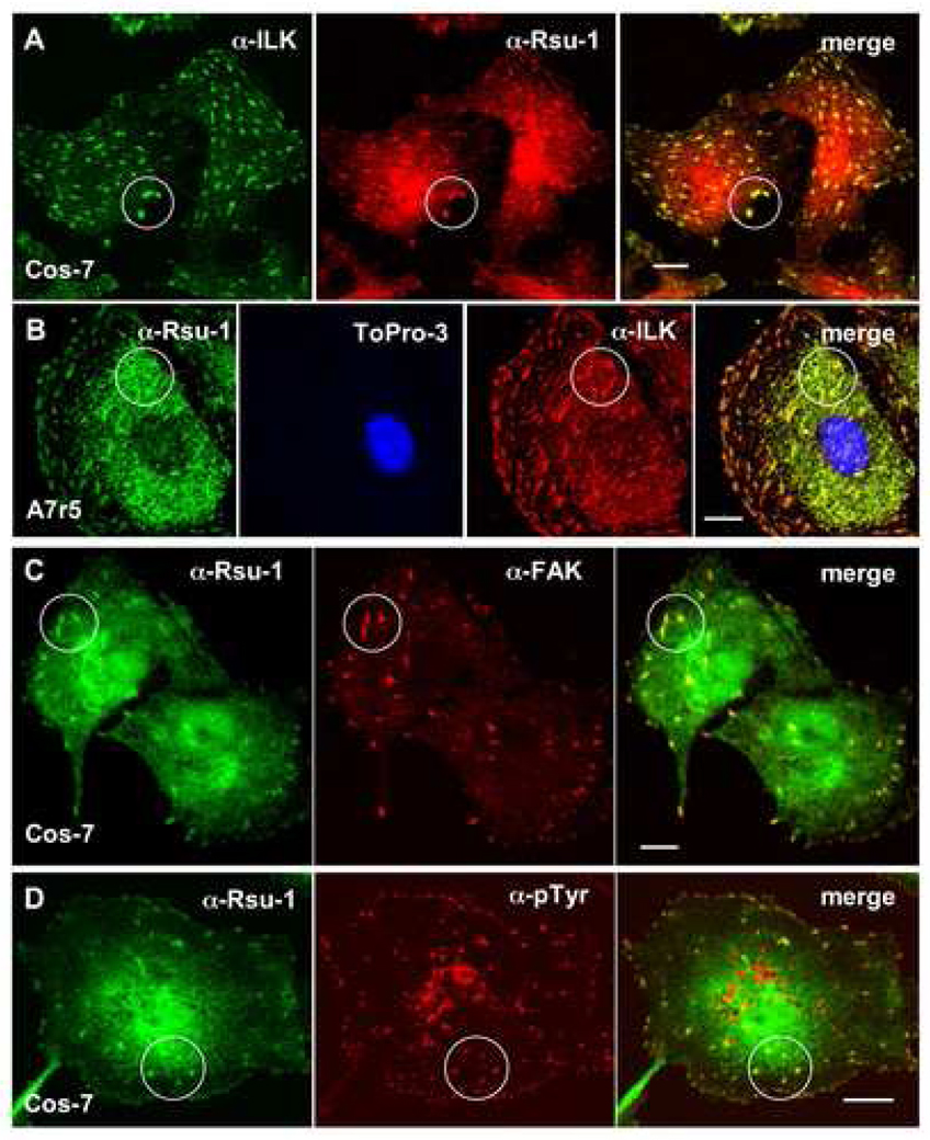
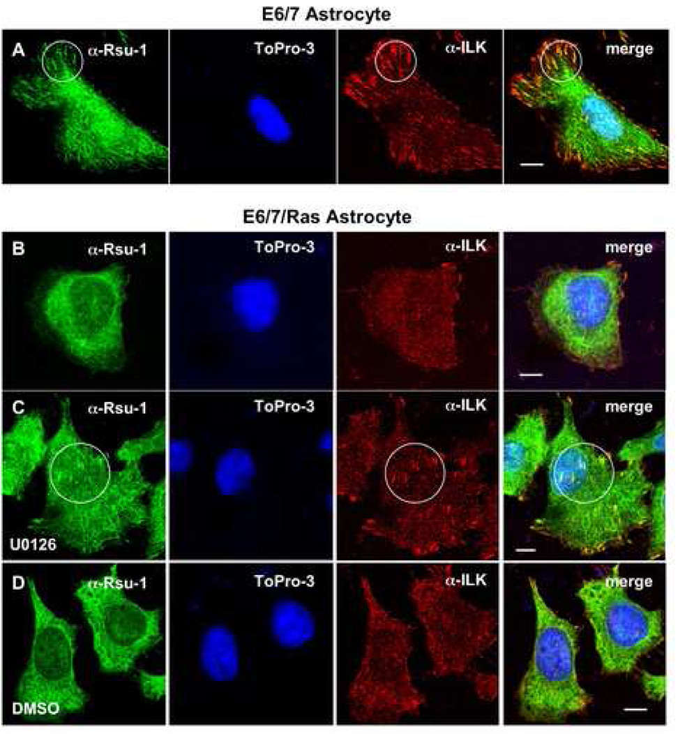
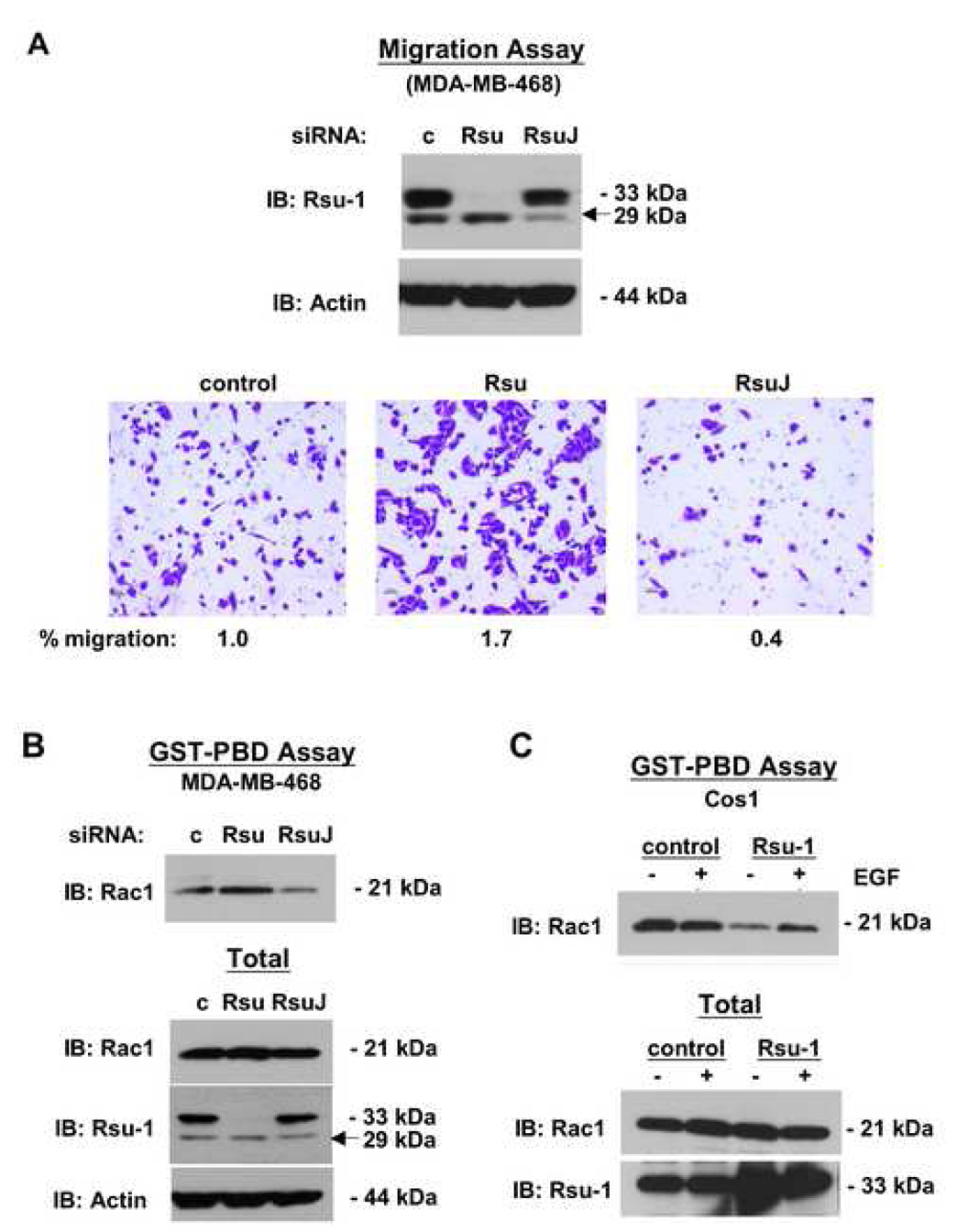
The link between Ras transformation and enhanced cell migration due to altered integrin signaling is well established in tumorigenesis, however there remain gaps in our understanding of its mechanism. The Ras suppressor, Rsu-1, has recently been linked to the IPP (integrin-linked kinase {ILK}, PINCH-1/LIMS1, parvin) focal adhesion complex based on its interaction with the LIM 5 domain of PINCH1. Defining the role of the Rsu1-PINCH1-ILK-parvin complex in tumorigenesis is important because both ILK and PINCH1 are elevated in certain tumors while ectopic expression of Rsu-1 blocks tumorigenesis. Our studies previously identified an alternatively spliced isoform of Rsu-1 in high-grade gliomas. We report here the detection of a truncated (p29) Rsu-1 protein, which correlates with the presence of the alternatively spliced Rsu-1 RNA. This RNA and the respective protein were detected in human tumor cell lines that contain high levels of activated Ras, and inhibitor studies demonstrate that the Mek-ERK pathway regulates expression of this truncated Rsu-1 product. We also show that Rsu-1 co-localizes with ILK at focal contacts and co-immunoprecipitates with the ILK-PINCH1 complex in non-transformed cells, but following Ras transformation the association of Rsu-1 with the PINCH1-ILK complex is greatly reduced. Using a human breast cancer cell line, our in vitro studies demonstrate that the depletion of Rsu-1 full-length protein enhances cell migration coincident with an increase in Rac-GTP while the depletion of the p29 Rsu-1 truncated protein inhibits migration. These findings indicate that Rsu-1 may inhibit cell migration by stabilizing the IPP adhesion complex and that Ras activation perturbs this inhibitory function by modulating both Rsu-1 splicing and association of full-length Rsu-1 with IPP. Hence, our findings demonstrate that Rsu-1 links the Ras pathway with the IPP complex and the perturbations of cell attachment-dependent signaling that occur in the malignant process.


Figures

Fig. 1
Fig. 1. Ras-activated human tumor cell lines and immortalized human astrocytes express a truncated 29-kDa Rsu-1 protein
(A) Lysates (100 µg in RIPA buffer) of human breast tumor cell lines were examined for expression of Rsu-1, PINCH1, ILK, and β-actin by Western blotting. Detection of actin was used as a loading control. Ras activation correlates with expression of a truncated 29-kDa Rsu-1 protein (arrow). (B) Cell lines expressing the truncated form of Rsu-1 contain high levels of Ras-GTP. Breast cancer cell lines were serum starved for 24 h and then stimulated with EGF for 7.5 min or left untreated. The level of Ras-GTP was determined by binding 1 mg lysate proteins to GST-Raf-RBD beads. The Ras-GTP bound to the beads and the total Ras protein in the lysates were detected by Western blotting using anti-pan Ras antibody. (C) Lysates (100 µg in RIPA buffer) of immortalized human astrocytes and their Ras-transformed counterparts were examined for expression of Rsu-1, PINCH1, ILK, and β-actin by Western blotting. Ras activation correlates with expression of a truncated 29-kDa Rsu-1 protein (arrow). (D) An alternatively-spliced transcript of Rsu-1 is detected in human breast cancer cell lines and Ras-transformed astrocytes; 1 µg RNA was used for RT-PCR to amplify the Rsu-1 open reading frame-specific sequence. RT-PCR products were separated on 1% agarose gels, stained with ethidium bromide and transferred to filters for Southern blotting with an Rsu-1 open reading frame-specific probe. A 725-bp product that encodes the p29 Rsu-1 protein is specifically detected in cell lines with Ras activation but not in matched controls.
Fig. 2
Fig. 2. The association of Rsu-1 with the PINCH1-ILK complex is altered in tumor cell lines
(A) Lysates of human breast cancer cell lines were immunoprecipitated with anti-PINCH1. The immunoprecipitates were analyzed by Western blotting for co-immunoprecipitation of endogenous Rsu-1. Only p33 Rsu-1 is detected in PINCH1 immunoprecipitates. (B) The Ras-transformed immortalized human astrocytes were treated with inhibitors at the indicated concentrations for 36 h and the effect on p33 and p29 Rsu-1 protein expression was determined by Western blotting. Inhibitors: 10 µM PD98059, 10 and 20 µM U0126 (left and right lanes, respectively), 10 µM LY29402, 500 nM SB20350, 100 nM JNKII (SP600125), 100 nM RhoK inhibitor (Y27632), 5 ng/ml TGFβ. (C) Lysates of immortalized human astrocytes and the Ras-transformed astrocyte cell line were immunoprecipitated with anti-ILK. The immunoprecipitates were analyzed by Western blotting for co-immunoprecipitation of endogenous Rsu-1 and PINCH1. (D) The ILK-immunoprecipitates of Ras-transformed astrocytes were analyzed by Western blotting for co-immunoprecipitation of endogenous Rsu-1 with and without pre-treatment of the cells with U0126.
Fig. 3
Fig. 3. Rsu-1 colocalizes with focal adhesion components
Cos-7 (A, C, D) or A7r5 (B) cells were plated on fibronectin-coated coverslips and assayed by immunofluorescence using anti-amino-terminal Rsu-1 (A–D), anti-ILK (A, B), anti-FAK (C), and anti-pTyr antibodies (D). Colocalizations are highlighted in circles. Nuclei were counterstained with Topro-3 in (B). Bars: 10 µm (A–D).
Fig. 4
Fig. 4. Ras transformation reduces Rsu-1 colocalization with ILK at focal adhesions
E6/7 astrocytes (A) and E6/7 astrocytes transformed with activated Ras (E6/7/Ras) (B–D) were plated on fibronectin-coated coverslips and assayed by immunofluorescence using anti-amino-terminal Rsu-1 and anti-ILK antibodies. Nuclei were counterstained with Topro-3 (to exclude cells with aberrant or multiple nuclei.). Colocalizations are highlighted in circles. Colocalization at focal adhesions is reduced in E6/7/Ras (B). Inhibition of ERK activation (20 µM U0126 for 24 h prior to plating) partially restores Rsu-1 and ILK colocalization at focal adhesions (C). (D) Solvent control (DMSO). Bars: 10 µm (A–D).
Fig. 5
Fig. 5. Effect of Rsu-1 expression on cell migration and levels of Rac-GTP
(A, B) MDA-MB-468 cells were depleted of p33 Rsu-1 (siRNA Rsu) or p29 Rsu-1 (siRNA RsuJ) for 72 h. (A) siRNA-treated cells (5 × 10⁴) were seeded on matrigel-coated membranes in Boyden chambers. At 20 h post seeding the cells migrating through the membrane were fixed, stained and enumerated (lower panels). The percentage of cells migrating was normalized to that of control siRNA-treated cells (siRNA c) which was set at 1.0. Upper panels: Verification of reduced Rsu-1 expression after siRNA treatment. (B) siRNA-treated cells were harvested and the level of Rac-GTP was determined by binding to the GST-Rac-binding domain of Pak1 (GST-PBD) (upper panel). (C) Cos-1 cells were transfected with an empty vector or a vector encoding p33 Rsu-1. At 72 h post transfection the cells lysates were prepared from cells with or without EGF stimulation (100 ng/ml, 7 min), and the level of Rac-GTP was determined (upper panel). Five percent of the total cell lysate was included as control for cellular levels of Rac (B, C; lower panels).

References

    1. Abassi YA, Vuori K. Tyrosine 221 in Crk regulates adhesion-dependent membrane localization of Crk and Rac and activation of Rac signaling. EMBO J. 2002;21:4571–4582. - PMC - PubMed
    1. Ahmed N, Riley C, Oliva K, Stutt E, Rice GE, Quinn MA. Integrin-linked kinase expression increases with ovarian tumour grade and is sustained by peritoneal tumour fluid. J. Pathol. 2003;201:229–237. - PubMed
    1. Albini A, Iwamoto Y, Kleinman HK, Martin GR, Aaronson SA, Kozlowski JM, McEwan RN. A rapid in vitro assay for quantitating the invasive potential of tumor cells. Cancer Res. 1987;47:3239–3245. - PubMed
    1. Almeida EA, Ilic D, Han Q, Hauck CR, Jin F, Kawakatsu H, Schlaepfer DD, Damsky CH. Matrix survival signaling: from fibronectin via focal adhesion kinase to c-Jun NH(2)-terminal kinase. J. Cell Biol. 2000;149:741–754. - PMC - PubMed
    1. Becker E, Huynh-Do U, Holland S, Pawson T, Daniel TO, Skolnik EY. Nck-interacting Ste20 kinase couples Eph receptors to c-Jun N-terminal kinase and integrin activation. Mol. Cell. Biol. 2000;20:1537–1545. - PMC - PubMed

Publication types

MeSH terms

LinkOut - more resources