

Comparing contrast-enhanced color flow imaging and pathological measures of breast lesion vascularity
- PMID: 18436369
- PMCID: PMC2556965
- DOI: 10.1016/j.ultrasmedbio.2008.02.010
Comparing contrast-enhanced color flow imaging and pathological measures of breast lesion vascularity
Abstract
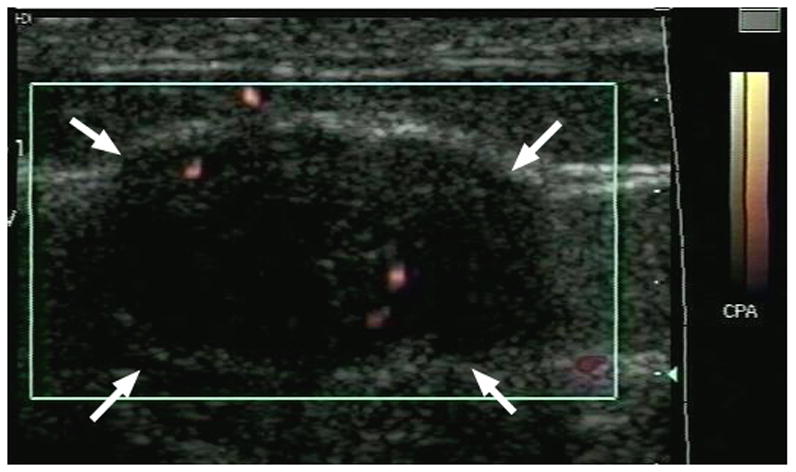
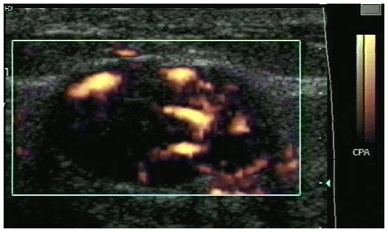
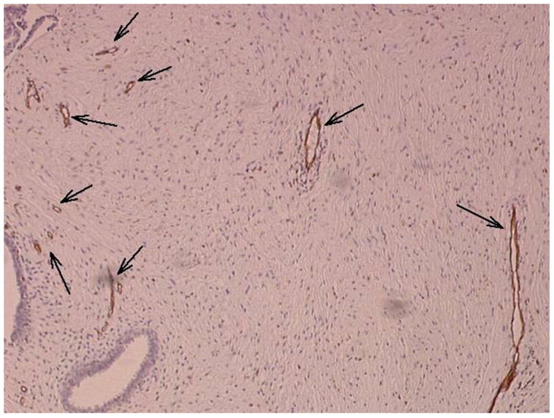
This study was conducted to compare quantifiable measures of vascularity obtained from contrast-enhanced color flow images of breast lesions to pathologic vascularity measurements. Nineteen patients with solid breast masses received Levovist Injection (10 mL at 300 mg/mL; Berlex Laboratories, Montville, NJ, USA). Color flow images of the mass pre and post contrast were obtained using an HDI 3000 scanner (Philips Medical Systems, Bothell, WA, USA) optimized for clinical scanning on an individual basis. After surgical removal, specimens were sectioned in the same planes as the ultrasound images and stained with an endothelial cell marker (CD31). Microvessel area (MVA) and intratumoral microvessel density (MVD) were determined for vessels 10-19 microm, 20-29 microm, 30-39 microm, 40-49 microm and > or =50 microm in diameter using a microscope and image processing software. From the ultrasound images, the number of color pixels before and after contrast administration relative to the total area of the breast mass was calculated as a first-order measure of fractional tumor vascularity. Vascularity measures were compared using reverse stepwise multiple linear regression analysis. In total, 58 pathology slides (with 8,106 frames) and 185 ultrasound images were analyzed. There was a significant increase in flow visualization pre to post Levovist injection (p = 0.001), but no differences were found between the 11 benign and the eight malignant lesions (p > 0.35). Ultrasound vascularity measurements post contrast correlated significantly with pathology (0.15 < or = r2 < or = 0.46; p < 0.03). The 30-39 microm vessel range contributed most significantly to the MVD relationship (p < 0.001), whereas the MVA was mainly influenced by vessels 20-29 microm (p < 0.004). Precontrast ultrasound only correlated with pathology for relative MVA (r2 = 0.16; p = 0.01). In conclusion, contrast-enhanced color flow imaging provides a noninvasive measure of breast tumor neovascularity, corresponding mainly to vessels 20-39 microm in diameter, when used in a typical clinical setting.
Figures
References
-
- Adler DD, Carson PL, Rubin JM, Quinn-Reid D. Doppler ultrasound color flow imaging in the study of breast cancer: preliminary findings. Ultrasound Med Biol. 1990;16:553–559. - PubMed
-
- Barbareschi M, Weidner N, Gasparini G, Morelli L, Forti S, Eccher C, Fina P, Caffo O, Leonardi E, Mauri F, Bevilacqua P, Palma PD. Microvessel density quantification in breast carcinomas: assessment by manual vs. a computer assisted image analysis system. Appl Immunohistochem. 1995;3:75–84.
-
- Bell DS, Bamber JC, Eckersley RJ. Segmentation and analysis of colour Doppler images of tumour vasculature. Ultrasound Med Biol. 1995;21:635–637. - PubMed
-
- Bohm-Velez M, Mendelson EB. Computed tomography, duplex Doppler ultrasound and magnetic resonance imaging in evaluating the breast. Semin Ultrasound CT MR. 1989;10:171–176. - PubMed
Publication types
MeSH terms
Substances
Grants and funding
LinkOut - more resources
Full Text Sources
Medical
