. 2008 Jun 20;283(25):17721-30.
doi: 10.1074/jbc.M800013200. Epub 2008 Apr 23.

Hydrogen peroxide promotes Abeta production through JNK-dependent activation of gamma-secretase

Affiliations

Hydrogen peroxide promotes Abeta production through JNK-dependent activation of gamma-secretase

Chengyong Shen et al. J Biol Chem. .

Abstract

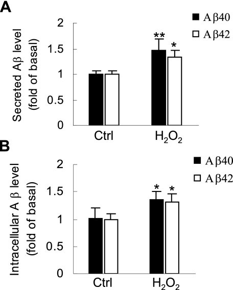
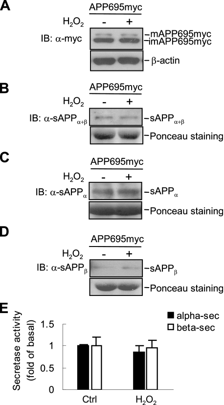
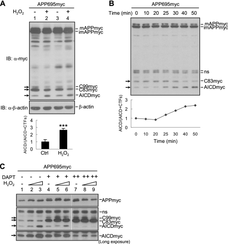
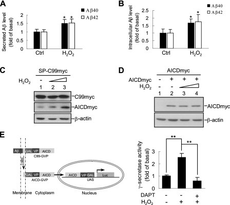
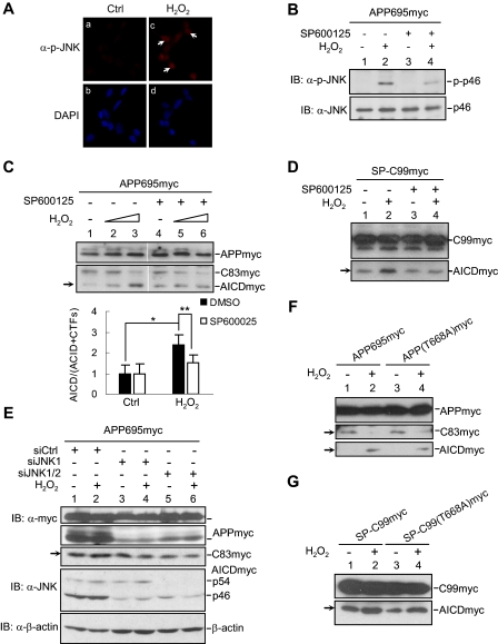
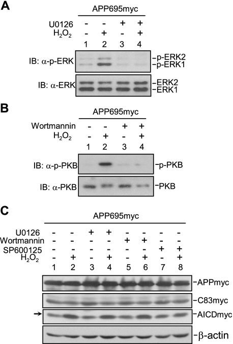
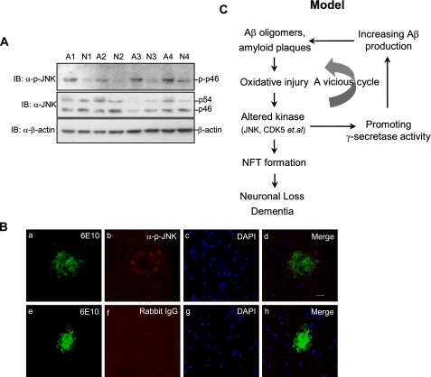
Accumulation of senile plaques composed of amyloid beta-peptide (Abeta) is a pathological hallmark of Alzheimer disease (AD), and Abeta is generated through the sequential cleavage of amyloid precursor protein (APP) by beta- and gamma-secretase. Although oxidative stress has been implicated in the AD pathogenesis by inducing Abeta production, the underlying mechanism remains elusive. Here we show that the pro-oxidant H(2)O(2) promotes Abeta production through c-Jun N-terminal kinase (JNK)-dependent activation of gamma-secretase. Treatment with H(2)O(2) induced significant increase in the levels of intracellular and secreted Abeta in human neuroblastoma SH-SY5Y cells. Although gamma-secretase-mediated cleavage of APP or C99 was enhanced upon H(2)O(2) treatment, expression of APP or its alpha/beta-secretase-mediated cleavage was not affected. Silencing of the stress-activated JNK by small interfering RNA or the specific JNK inhibitor SP600125 reduced H(2)O(2)-induced gamma-secretase-mediated cleavage of APP. JNK activity was augmented in human brain tissues from AD patients and active JNK located surrounding the senile plaques in the brain of AD model mouse. Our data suggest that oxidative stress-activated JNK may contribute to senile plaque expansion through the promotion of gamma-secretase-mediated APP cleavage and Abeta production.


Figures

FIGURE 1.
FIGURE 1.
H2O2 significantly induces intracellular and secreted Aβ. A, SH-SY5Y/APP695myc cells were treated with 1.0 mm H2O2 in serum-free medium for 1 h or left untreated as indicated. Conditioned medium was collected and analyzed by sandwich Aβ ELISA to measure secreted Aβ40 and Aβ42 level. B, SH-SY5Y/APP695myc cells were lysed in ice-cold radioimmune precipitation assay buffer, and the same cell extracts were subjected to Aβ ELISA. Ctrl, control.
FIGURE 2.
FIGURE 2.
H2O2 does not induce APP expression or stimulate the activity of α- and β-secretase. A, after H2O2 treatment, SH-SY5Y/APP695myc cells were analyzed by immunoblotting (IB) using anti-Myc antibody. B-D, conditioned medium was collected and analyzed by immunoblotting, using 22C11 antibody to detect total sAPP (B), 6E10 antibody to detect sAPPα (C), and anti-sAPPβ antibody to detect sAPPβ (D). Loading controls (Ctrl) of conditioned medium were analyzed by Ponceau staining. E, determination of α- and β-secretase activity by fluorogenic substrate assays. Lysates of H2O2-treated cells were incubated with secretase substances at 37 °C for 2 h, and emission was measured.
FIGURE 3.
FIGURE 3.
H2O2 promotes γ-secretase-mediated processing of APP. A, SH-SY5Y/APP695myc cells were treated with 1.0 mm H2O2, followed by immunoblotting (IB) using anti-Myc antibody. Quantification of AICD and CTFs was performed through normalization of APP level (bottom panel). B, a time course of H2O2-induced γ-secretase-mediated APP processing. The cells were treated with H2O2 up to 50 min and harvested at indicated time points. APP processing was analyzed as described in A. C, cells were pretreated with the γ-secretase inhibitor DAPT (1 and 10 μm) or the control Me2SO for 3 h, followed by H2O2 treatment. APP processing was monitored by immunoblotting as described in A. ns, nonspecific.
FIGURE 4.
FIGURE 4.
H2O2 promotesγ-secretase-mediated SP-C99myc processing. A, determination of H2O2-induced secreted Aβ40 and Aβ42 production. HEK293T cells were transiently transfected with pcDNA3.1-SP-C99myc for 24 h. Conditioned medium of H2O2-treated cells was collected and analyzed by ELISA to measure secreted Aβ40 and Aβ42 levels. B, determination of intracellular Aβ40 and Aβ42 levels in above cells. C, immunoblotting analysis of H2O2-induced AICD production in a dose-dependent manner in above cells (0.2 and 1.0 mm H2O2). D, HEK293T cells were transfected with pcDNA3.1-AICDmyc or empty vector and treated with various doses of H2O2 (0.2 and 1.0 mm H2O2). AICD accumulation was analyzed by immunoblotting using anti-Myc antibody. E, schematic representation of γ-secretase-dependent luciferase reporter assay (left panel). HEK293T cells were co-transfected with expression plasmids encoding C99-GVP, pFR-luc, and pRL-TK. The cells were pretreated with DAPT (10 μm) for 12 h, followed by H2O2 for another hour. Relative luciferase activity was analyzed as described under “Experimental Procedures” (right panel). Ctrl, control.
FIGURE 5.
FIGURE 5.
H2O2 promotes γ-secretase-mediated APP cleavage through JNK activation. A, immunofluorescent staining of JNK activation in H2O2-treated SH-SY5Y cells. B, immunoblot (IB) analysis of H2O2-induced JNK activation in SH-SY5Y cells. The cells were serum-starved for 12 h and pretreated with JNK inhibitor SP600125 (20 μm) or Me2SO control (Ctrl) for 3 h. After incubation with H2O2 for 15 min, the cells were harvested, and JNK activation was analyzed by immunoblotting with anti-phospho-JNK antibody. C, determination of the inhibitory effect of SP600125 on γ-secretase-mediated cleavage of APP695myc in H2O2-treated SH-SY5Y/APP695myc cells. D, determination of the inhibitory effect of SP600125 on γ-secretase-mediated cleavage in H2O2-treated HEK293T/SP-C99myc cells. E, the effect of siJNK on H2O2-induced γ-secretase-mediated APP cleavage. HEK293T cells were co-transfected with APP695myc and siRNA against Jnk1 or Jnk1/2. After H2O2 stimulation, the cells were analyzed by immunoblotting. F and G, immunoblot analysis of H2O2 effect on the processing of APP695myc(T668A) mutant (F) or SP-C99myc(T668A) mutant (G).
FIGURE 6.
FIGURE 6.
JNK, but not ERK or PKB, is required for H2O2 to promote γ-secretase-mediated APP cleavage. The cells were serum-starved for 12 h and pretreated with U0126 (5 μm) (A), wortmannin (20 nm) (B), SP600125 (20 μm) (C) or the control Me2SO for 3 h. After H2O2 treatment, the cells were analyzed by immunoblotting (IB) to determine the effect of the above inhibitors on activation of ERK (A), PKB (B), and APP processing (C).
FIGURE 7.
FIGURE 7.
JNK is activated in the brain areas that surround senile plaques in vivo. A, human brain samples (30 μg of total protein extracts) from AD (n = 4) and age-matched control samples (n = 4) were analyzed for JNK activation using anti-phospho-JNK and anti-pan JNK antibodies. B, immunostaining of active JNK and senile plaques in Tg2576 mouse brain. Sections were stained with 6E10 (green, panels a and e) and antiphospho-JNK antibody (red, panel b). Normal rabbit IgG was used as a negative control of anti-phospho-JNK (panel f). The nucleus was indicated with 4′,6′-diamino-2-phenylindole (DAPI, blue, panels c and g). The scale bar represents 20 μm. C, the model of H2O2 via JNK promotes γ-secretase-mediated APP processing. See “Discussion ” for the details. IB, immunoblotting.

References

    1. Selkoe, D. J. (2002) Science 298 789-791 - PubMed
    1. Saido, T. C., and Iwata, N. (2006) Neurosci. Res. 54 235-253 - PubMed
    1. Funato, H., Yoshimura, M., Kusui, K., Tamaoka, A., Ishikawa, K., Ohkoshi, N., Namekata, K., Okeda, R., and Ihara, Y. (1998) Am. J. Pathol. 152 1633-1640 - PMC - PubMed
    1. Mattson, M. P. (2004) Nature 430 631-639 - PMC - PubMed
    1. Selkoe, D. J., and Wolfe, M. S. (2007) Cell 131 215-221 - PubMed

Publication types