. 2008 Aug 1;79(3):395-404.
doi: 10.1093/cvr/cvn097. Epub 2008 Apr 23.

Colony-stimulating factor-1 transfection of myoblasts improves the repair of failing myocardium following autologous myoblast transplantation

Affiliations

Colony-stimulating factor-1 transfection of myoblasts improves the repair of failing myocardium following autologous myoblast transplantation

Seyedhossein Aharinejad et al. Cardiovasc Res. .

Abstract

Aims: Skeletal myoblasts are used in repair of ischaemic myocardium. However, a large fraction of grafted myoblasts degenerate upon engraftment. Colony-stimulating factor-1 (CSF-1) accelerates myoblast proliferation and angiogenesis. We hypothesized that CSF-1 overexpression improves myoblast survival and cardiac function in ischaemia-induced heart failure.

Methods and results: Three weeks following myocardial infarction, rats developed heart failure and received intramyocardial injections of mouse CSF-1-transfected or untransfected primary autologous rat myoblasts, recombinant human CSF-1, mouse CSF-1 expressing plasmids, or culture medium. Tissue gene and protein expression was measured by quantitative RT-PCR (reverse transcription-polymerase chain reaction) and western blotting. Fluorescence imaging and immunocytochemistry were used to analyse myoblasts, endothelial cells, macrophages, and infarct wall thickening. Electrocardiograms were recorded online using a telemetry system. Left ventricular function was assessed by echocardiography over time, and improved significantly only in the CSF-1-overexpressing myoblast group. CSF-1-overexpression enhanced myoblast numbers and was associated with an increased infarct wall thickness, enhanced angiogenesis, increased macrophage recruitment and upregulated matrix metalloproteases (MMP)-2 and -12 in the zone bordering the infarction. Transplantation of CSF-1-overexpressing myoblasts did not result in major arrhythmias.

Conclusion: Autologous intramyocardial transplantation of CSF-1 overexpressing myoblasts might be a novel strategy in the treatment of ischaemia-induced heart failure.


Figures

Figure 1
Figure 1
Functional analysis of hearts. Left ventricular ejection fraction (LVEF; A) and left ventricular end-diastolic volume (LVEDV; B) within each group at different time points. Only colony-stimulating factor-1 (CSF-1)-transfected myoblasts (MB-CSF-1) improved cardiac LVEF and LVEDV significantly over time, while LVEF and LVEDV of untreated myoblasts (MB), the recombinant human CSF-1 (recCSF-1), CSF-1 expression plasmid (pl-CSF-1), and untreated control groups vs. sham operated rats (Sham) had lesser or no effects. Data are expressed as means ± SD. (Asterisk) significantly different from sham on days (d) 7, 52, and 86, respectively; A: P < 0.001; B: P < 0.001 for controls, MB, recCSF-1; P < 0.001, P < 0.001, and P = 0.004 for pl-CSF-1; and P < 0.001 and P = 0.004 vs. sham group on d7 and d52, respectively, for MB-CSF-1. (Dagger) significantly different within an individual group; A: P < 0.001 and P = 0.007, MB-CSF-1 vs. d7 and d52, respectively; B: P < 0.001, Control d52 vs. d7; P < 0.001 and P = 0.004, MB d52 and d86, respectively, vs. d7; P < 0.001, recCSF-1 d52 vs. d7; P < 0.001, pl-CSF-1 d52 vs. d7. (Double dagger) significantly different from MB (A: P < 0.001 for d52, and P < 0.001 for d86 vs. d52, and d86; B: P = 0.007 for d52, and P < 0.005 for d86 vs. d52 and d86.
Figure 2
Figure 2
Morphometric analysis of hearts. Representative images (A) and quantitative morphometric analysis (B) of collagenous scar tissue in left ventricular (LV) myocardium by Goldner trichrome stain on day 88 after infarction in the control, myoblasts (MB), the recombinant human colony-stimulating factor-1 (recCSF-1), CSF-1 expression plasmid (pl-CSF-1), and CSF-1-transfected myoblasts (MB-CSF-1) groups. LV infarct wall thickness (C) and heart-to-body weight ratio (D). Data are expressed as means ± SD. (Asterisk) significantly different from sham (C: P < 0.001; D: P = 0.002, P = 0.012, P = 0.003, and P = 0.029 for controls, MB, recCSF-1, and pl-CSF-1, respectively); (Dagger) significantly different from control, MB, recCSF-1, and pl-CSF-1 (P < 0.014).
Figure 3
Figure 3
Localization of DiIC18-labelled autologous rat myoblasts (DiI-MB) and DiIC18-labelled colony-stimulating factor-1 (CSF-1)-overexpressing autologous rat myoblasts (DiI MB-CSF-1) in the heart, obtained 88 days after infarction. (A) Representative white-light images (left), colour-coded fluorescent map of DiIC18-signal (middle), and image overlays of the heart produced in Adobe Photoshop (right) showing the localization of DiIC18-labelled myoblasts (red/yellow) in the infarction area of the heart (Asterisk). The fluorescence signals from the implanted myoblasts in the heart are strongest in the DiI MB-CSF-1 group. LV, left ventricle; RV, right ventricle; the atriums are removed. (B) Representative red-channel fluorescence images of DiIC18-signal (left), image overlay of the red fluorescence images with the corresponding section counterstained with DAPI (middle) and HE-staining of the same section (right) in the border zone of the DiI-MB and DiI MB-CSF-1 groups. The HE stain (right) defines the morphology in the border zone of the infarction area with integrated myoblasts. The myoblasts are easily identified on the fluorescent optical images from the surrounding tissue, because of their bright fluorescence. (C) Representative images of myocytes stained by skeletal muscle myosin heavy chain specific antibody in both the MB (upper image) and MB-CSF-1 (lower image) groups, showing integration of transplanted myoblasts (arrows) into neighboring myocardial tissue Bar = 50 µm. (D) Quantitative analysis of DiIC18-labelled grafted myoblasts in the heart. Data are expressed as means ± SD. (Asterisk) significantly different from control and MB (P < 0.02). (E) CSF-1 gene transfer increases proliferation of myoblasts in vitro. Values are expressed as means ± SD. (Asterisk) significantly different from MB (P = 0.003) at 72 h. (F) PCNA stain (green) of DiIC18-labelled (red) MB and MB-CSF-1 transplanted groups. PCNA-positive MBs are not detected. Occasional PCNA positive proliferative events of non-MB cells are detected (insert).
Figure 4
Figure 4
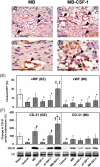
Analysis of angiogenesis. Representative vWF-stained sections in the myoblasts (MB) (A, B) and MB-colony-stimulating factor-1 (CSF-1) (C, D) groups, reveal a large number of vessels (arrows) in areas with transplanted myoblasts. Bar = 50 µm. Quantification of vWF-positive vessels in myocardial tissue of the border zone (BZ) and the myocardial infarction zone (MI) 88 days after infarction per high-power field (HPF; E). Representative western blot images and quantification of protein levels of CD-31 western blots in BZ and the MI zone 88 days after infarction. Actin was used as a loading control (LC) (F). Data are expressed as means ± SD. (Asterisk) significantly different vs. sham (E: P < 0.025; F: P < 0.001); (Dagger) significantly different vs. control [E: P = 0.001; F: P < 0.001 for MB and MB-CSF-1, respectively, and P = 0.01 for CSF-1 expression plasmid (pl-CSF-1)]; (Double dagger) significantly different from MB, recombinant human CSF-1 (recCSF-1), and pl-CSF-1 (P < 0.001).
Figure 5
Figure 5
Analysis of colony-stimulating factor-1 (CSF-1) expression and macrophages. (A) CSF-1 mRNA expression levels following transfection of myoblasts with CSF-1 expression plasmids in vitro, normalized to GAPDH. (Asterisk) significantly different from control P < 0.001). (B) Analysis of myocardial CSF-1 mRNA expression on days (d) 52 and 88 after myocardial infarction normalized to GAPDH. Data are expressed as means ± SD. (Asterisk) significantly different from other groups (P < 0.05). (C) Quantitative histo-morphometric analysis of myocardial tissue sections of the border zone on d88 after infarction stained with the macrophage specific RM-4 antibody. Data are expressed as means ± SD. (Asterisk) significantly different from control (A: P < 0.001; B: P = 0.047). Representative immunocytochemistry images of myocardial tissue sections of the border zone on d88 after infarction in MB (D, E) and MB-CSF-1 (F, G) group stained with the macrophage specific RM-4 antibody. Arrowheads indicate macrophages stained positively with RM-4 antibody inside the myocardial tissue. Calibration bar = 50 µm.
Figure 6
Figure 6
Analysis of matrix metalloproteases (MMP) expression. (A) Quantification of protein expression levels of MMP-2, MMP-9, MMP-1, MMP-13, and MMP-12 and representative western blot images in myocardial tissue of the border zone (BZ) and the myocardial infarction zone (MI) 88 days after myocardial infarction. Actin was used as a loading control (LC); SP, specific protein. Data are expressed as means ± SD. MMP-2: (Asterisk) significantly different from control (P = 0.006); MMP-9: (Asterisk) significantly different from control [P < 0.0001 for myoblasts (MB) group; P = 0.001 for MB-colony-stimulating factor-1 (CSF-1) group]; MMP-13: (Asterisk) significantly different from control (P = 0.045 for MB group; P = 0.003 for MB-CSF-1 group); MMP-12: (Asterisk) significantly different from control (P = 0.039). (B) Representative immunocytochemistry images of MMP-2- (left panels) and MMP-12- (right panels) stained myocardial sections in MB (upper panels) and MB-CSF-1 (lower panels). Both MMP-2- and MMP-12-positive cells (arrows) reveal a higher density in the MB-CSF-1 group. Bar = 50 µm.

Comment in

References

    1. Menasche P, Hagege AA, Vilquin JT, Desnos M, Abergel E, Pouzet B, et al. Autologous skeletal myoblast transplantation for severe postinfarction left ventricular dysfunction. J Am Coll Cardiol. 2003;41:1078–1083. - PubMed
    1. Murry CE, Wiseman RW, Schwartz SM, Hauschka SD. Skeletal myoblast transplantation for repair of myocardial necrosis. J Clin Invest. 1996;98:2512–2523. - PMC - PubMed
    1. Taylor DA, Atkins BZ, Hungspreugs P, Jones TR, Reedy MC, Hutcheson KA, et al. Regenerating functional myocardium: improved performance after skeletal myoblast transplantation. Nat Med. 1998;4:929–933. - PubMed
    1. Jain M, DerSimonian H, Brenner DA, Ngoy S, Teller P, Edge AS, et al. Cell therapy attenuates deleterious ventricular remodeling and improves cardiac performance after myocardial infarction. Circulation. 2001;103:1920–1927. - PubMed
    1. Menasche P, Hagege AA, Scorsin M, Pouzet B, Desnos M, Duboc D, et al. Myoblast transplantation for heart failure. Lancet. 2001;357:279–280. - PubMed

Publication types

MeSH terms

Substances