. 2008 Jul;84(1):170-81.
doi: 10.1189/jlb.0208100. Epub 2008 Apr 24.

The acute phase protein haptoglobin regulates host immunity

Affiliations

The acute phase protein haptoglobin regulates host immunity

Kristin M Huntoon et al. J Leukoc Biol. 2008 Jul.

Abstract

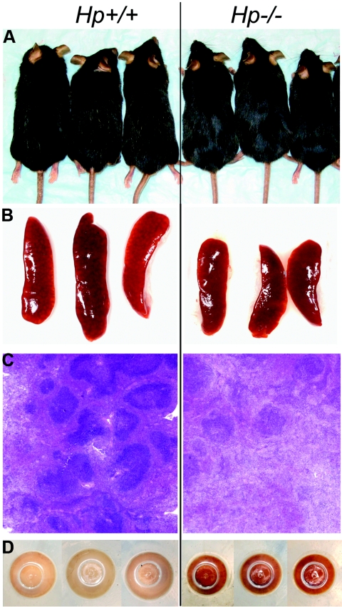
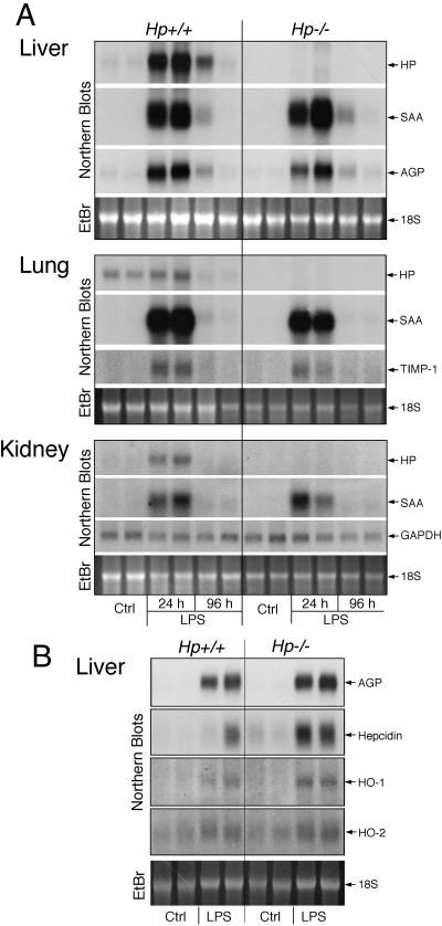
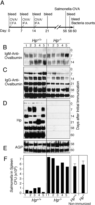
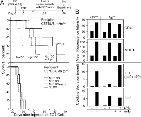
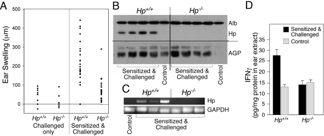
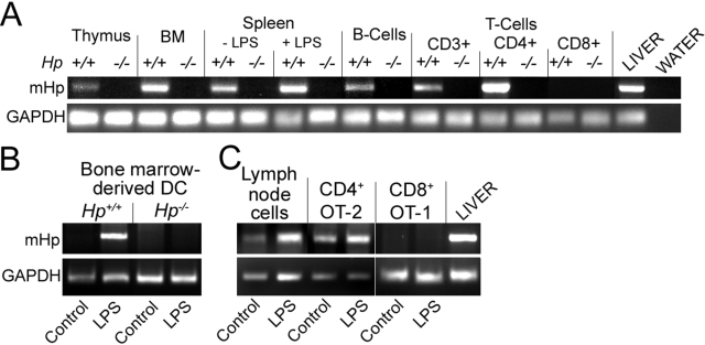
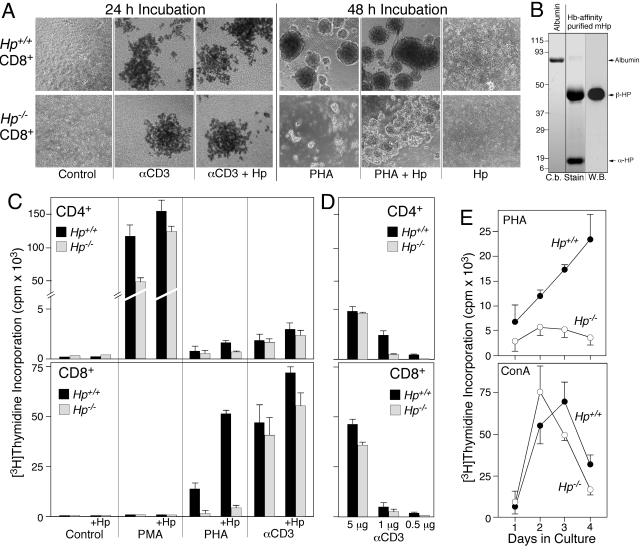
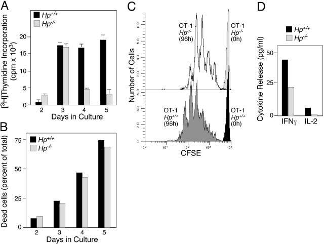
The contribution of acute phase plasma proteins to host immune responses remains poorly characterized. To better understand the role of the acute phase reactant and major hemoglobin-binding protein haptoglobin (Hp) on the function of immune cells, we generated Hp-deficient C57BL/6J mice. These mice exhibit stunted development of lymphoid organs associated with lower counts of mature T and B cells in the blood and secondary lymphoid compartments. Moreover, these mice show markedly reduced adaptive immune responses as represented by reduced accumulation of IgG antibody after immunization with adjuvant and nominal antigen, abrogation of Th1-dominated delayed-type hypersensitivity reaction, loss of mitogenic responses mounted by T cells, and reduced T cell responses conveyed by APCs. Collectively, these defects are in agreement with the observations that Hp-deficient mice are not capable of generating a recall response or deterring a Salmonella infection as well as failing to generate tumor antigen-specific responses. The administration of Hp to lymphocytes in tissue culture partially ameliorates these functional defects, lending further support to our contention that the acute phase response protein Hp has the ability to regulate immune cell responses and host immunity. The phenotype of Hp-deficient mice suggests a major regulatory activity for Hp in supporting proliferation and functional differentiation of B and T cells as part of homeostasis and in response to antigen stimulation.


Figures

Figure 1
Figure 1
Phenotypic markers for Hp deficiency in C57BL/6J mice. Comparison of age-matched (8-month-old) Hp+/+ and Hp−/− mice: (A) black and shining hair; (B) smaller spleen size; (C) lower number and smaller size germinal centers in spleen (H&E staining); and (D) presence of Hb precipitates within splenocytes (bottom view of the pellet of isolated cells in centrifuge tube).
Figure 2
Figure 2
LPS-mediated induction of acute phase reactants in various organs is maintained in Hp−/− mice. (A) Age-matched (10-week-old) Hp+/+ and Hp−/− C57BL/6J mice received a single i.p. injection of PBS (Ctrl) or 50 μg LPS, and after 24 h (Ctrl and LPS) or 96 h (LPS), RNAs were extracted from the indicated organs of two animals and analyzed by Northern blotting for the mRNA listed on the right. (B) A separate set of 10-week-old animals received PBS (Ctrl) or 50 μg LPS, and 24 h later, liver RNAs were isolated and analyzed by Northern blotting for the indicated mRNAs. SAA, Serum amyloid A; EtBr, ethidium bromide; TIMP-1, tissue inhibitor for metalloproteinase-1; HO, heme oxygenase.
Figure 3
Figure 3
Hp deficiency attenuates the immune response to OVA. (A) Schematic time-line of the experiment, indicating the sequence of bleedings and immunization with OVA in CFA or in IFA and final challenge with OVA-expressing Salmonella. (B–F) Groups of five age-matched (8-week-old) Hp+/+ and Hp−/− mice were immunized with OVA. Aliquots of the blood taken at the days indicated were analyzed by immunoblotting for the level of OVA-specific IgM (B), IgG (C), Hp (D), and AGP (E). Titer of Salmonella-OVA in the spleen extracts 4 days after challenge was determined. A significant difference was observed between Hp-deficient animals and wild-type (P=0.0002). Salmonella growth in nonimmunized animals served as reference (mean±se, n=5; F). Results are representative of two independent experiments with five mice in each group.
Figure 4
Figure 4
Effectiveness of immunization by DC from Hp+/+ and Hp−/− mice and influence of systemic Hp. (A) Groups of 5 Hp+/+ and Hp−/− C57BL/6 mice received 1 × 10⁶ matured bone marrow-derived DC from C57BL/6J.Hp+/+ or Hp−/− mice (Day –5). Prior to transfer, the DC had been treated for 4 h with OVA, followed by 16 h with LPS. One culture with Hp−/− DC also included 1 mg/ml mouse Hp. The recipient mice were challenged i.p. with 1 × 10⁶ E.G7 (EL4 OVA-expressing thymoma) tumor cells (=Day 0). Mice were then monitored for survival. In Hp+/+ mice, Hp+/+ DC versus Hp−/− DC: P = 0.01; Hp−/− DC + mouse Hp (mHp) versus Hp−/− DC: P = 0.02. (B) Bone marrow-derived DC from the indicated genotypes (1×10⁶ cells/ml) were treated for 24 h with LPS (1 μg/ml) or Hp (1 mg/ml). The expression of cell surface markers was identified by flow cytometry, and the cytokine concentrations in the conditioned medium were determined by Luminex immunobead assay. Data represent the results from one experimental series. Although secretion rate varies among independently derived cell preparations, in three series, an identical trend in Hp-dependent cytokine regulation was determined.
Figure 5
Figure 5
Hp deficiency suppresses a DTH reaction. (A) Ear swelling was determined on Hp+/+ or Hp−/− mice 24 h after challenge with DNFB as a function of phenotype and prior sensitization. (B) Western blot analysis ear lysates from Hp+/+ and Hp−/− mice sensitized and challenged indicate the presence of Hp and AGP proteins. Albumin (Alb) is shown to demonstrate equal loading of serum proteins. Each lane represents an independent, individual mouse. Lanes labeled as “Control” represent extracts of animals with challenge only. (C) RNAs extracted from ear tissues from Hp+/+ or Hp−/− mice 24 h after a challenge were determined by RT-PCR for the expression of Hp and GAPDH mRNAs. Each lane represents an independent, individual mouse. (D) Extracts from ear tissue from Hp+/+ or Hp−/− mice 24 h after challenge were used to quantify the concentration of IFN-γ (means±sd, n=4).
Figure 6
Figure 6
Hp mRNA is expressed in lymphoid organs and in a subset of lymphoid cells. The indicated organs and hematopoietic cell types were prepared from 12-week-old mice. In selected cases, the animals received a LPS injection 24 h prior to organ collection. RNAs were extracted from intact organs after isolation of the cells (A), or from cell cultures treated in vitro for 24 h with 10 μg/ml LPS (B and C) and analyzed by RT-PCR for Hp and GAPDH. BM, Bone marrow.
Figure 7
Figure 7
T cells from Hp-deficient mice show an altered response to mitogens. T cells from Hp-deficient mice show an altered response to mitogens (A), microphotographs (4×) of purified CD8 cells isolated from Hp+/+ and Hp−/− C57BL/6J mice treated for 24 h with anti-αCD3 (αCD3), or for 48 h with PHA. Selected cultures also included 1 mg/ml mouse Hp. (B) Coomassie blue-stained SDS-PAGE gel (C.b. Stain) demonstrates purity of Hp (8 μg) isolated from acute phase serum of Hp+/+ C57BL/6J. A duplicate lane was used for Western blotting (W.B.) reacted with rabbit anti-Hp (recognizes only epitopes in the β-subunit). (C and D) [3H]Thymidine incorporation was determined in cultures of purified CD4 and CD8 cells that had been maintained in the presence of the indicated mitogen for 72 h. (E) DNA synthesis was determined in nonfractionated splenocytes from Hp+/+ and Hp−/− mice as a function of duration in culture in the presence of PHA or Con A.
Figure 8
Figure 8
Attenuated response of CD8+ T cells from Hp−/− mice. (A) CD8+ OT-1 cells were isolated from spleens of Hp+/+ or Hp−/− C57BL/6J background and cultured in the presence of irradiated BOK cells presenting H-2Kb-SIINFEKL and B7.1. DNA synthesis ([3H]Thymidine Incorporation) at Days 2–5 was determined. (B) The relative presence of apoptotic cells in the cultures at the times indicated was determined by flow cytometric analysis using labeling with Annexin V-FITC and PI. (C) CFSE-labeled, naive OT-1 T cells from Hp+/+ or Hp−/− at the onset and after 96 h stimulation by coculture with BOK cells were analyzed by flow cytometry. (D) OT-1 T cells from Hp+/+ or Hp−/− were stimulated by coculture with BOK cells for 72 h, and conditioned medium was analyzed for concentration of IFN-γ and IL-2.

References

    1. Jayle M. F., Boussier G., Badin J. Electrophoresis of haptoglobin and of its hemoglobin complex. Bull Soc Chim Biol (Paris) 1952;34:1063–1069. - PubMed
    1. Wicher K. B., Fries E. Haptoglobin, a hemoglobin-binding plasma protein, is present in bony fish and mammals but not in frog and chicken. Proc Natl Acad Sci USA. 2006;103:4168–4173. - PMC - PubMed
    1. Dobryszycka W. Biological functions of haptoglobin—new pieces to an old puzzle. Eur J Clin Chem Clin Biochem. 1997;35:647–654. - PubMed
    1. Wang Y., Kinzie E., Berger F. G., Lim S. K., Baumann H. Haptoglobin, an inflammation-inducible plasma protein. Redox Rep. 2001;6:379–385. - PubMed
    1. Oh S. K., Ross S., Walker J., Zeisel S. Role of a SER immune suppressor in immune surveillance. Immunology. 1988;64:73–79. - PMC - PubMed

Publication types

MeSH terms