. 2008 Jul;84(1):302-10.
doi: 10.1189/jlb.0208090. Epub 2008 Apr 24.

Neuropilin-1 is a receptor for transforming growth factor beta-1, activates its latent form, and promotes regulatory T cell activity

Affiliations

Neuropilin-1 is a receptor for transforming growth factor beta-1, activates its latent form, and promotes regulatory T cell activity

Yelena Glinka et al. J Leukoc Biol. 2008 Jul.

Abstract

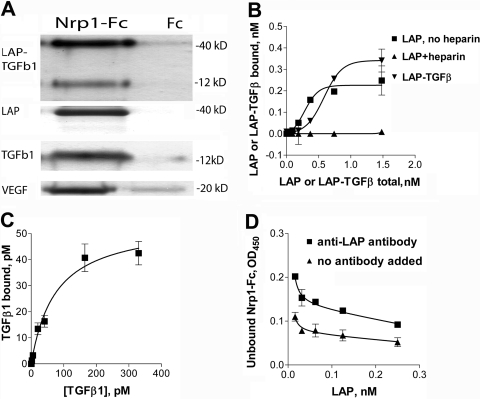
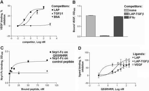
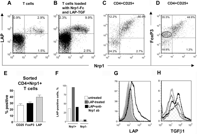
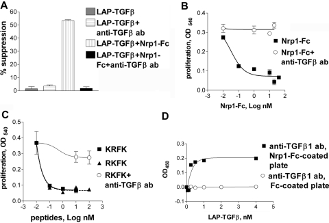
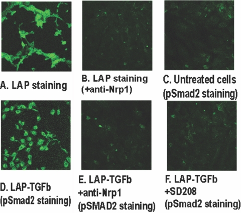
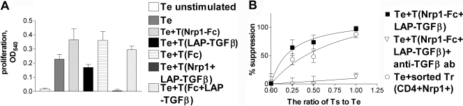
Neuropilin-1 (Nrp1) is a multifunctional protein, identified principally as a receptor for the class 3 semaphorins and members of the vascular endothelial growth factor (VEGF) family, but it is capable of other interactions. It is a marker of regulatory T cells (Tr), which often carry Nrp1 and latency-associated peptide (LAP)-TGF-beta1 (the latent form). The signaling TGF-beta1 receptors bind only active TGF-beta1, and we hypothesized that Nrp1 binds the latent form. Indeed, we found that Nrp1 is a high-affinity receptor for latent and active TGF-beta1. Free LAP, LAP-TGF-beta1, and active TGF-beta1 all competed with VEGF165 for binding to Nrp1. LAP has a basic, arginine-rich C-terminal motif similar to VEGF and peptides that bind to the b1 domain of Nrp1. A C-terminal LAP peptide (QSSRHRR) bound to Nrp1 and inhibited the binding of VEGF and LAP-TGF-beta1. We also analyzed the effects of Nrp1/LAP-TGF-beta1 coexpression on T cell function. Compared with Nrp1(-) cells, sorted Nrp1+ T cells had a much greater capacity to capture LAP-TGF-beta1. Sorted Nrp1(-) T cells captured soluble Nrp1-Fc, and this increased their ability to capture LAP-TGF-beta1. Conventional CD4+CD25(-)Nrp1(-) T cells coated with Nrp1-Fc/LAP-TGF-beta1 acquired strong Tr activity. Moreover, LAP-TGF-beta was activated by Nrp1-Fc and also by a peptide of the b2 domain of Nrp1 (RKFK; similar to a thrombospondin-1 peptide). Breast cancer cells, which express Nrp1, also captured and activated LAP-TGF-beta1 in a Nrp1-dependent manner. Thus, Nrp1 is a receptor for TGF-beta1, activates its latent form, and is relevant to Tr activity and tumor biology.


Figures

Fig. 1.
Fig. 1.
Nrp1 binds TGF-β1 components. (A) LAP-TGF-β1, LAP (β1), TGF-β1, and VEGF bound to Nrp1-Fc (but not control Fc) and were retained on protein G-sepharose beads. Bound proteins were recovered and immunoblots performed with specific antibodies. Molecular weight markers are indicated. (B) To demonstrate binding by ELISA, Nrp1-Fc-coated plates were incubated with increasing concentrations of the ligands. LAP (alone but not in the presence of 2 μg/ml heparin) and LAP-TGF-β1 bound at high affinity to Nrp1-Fc (see text). Several control proteins, including IFN-γ and IL-2, did not bind (not shown). (C) Active TGF-β1 bound to immobilized Nrp1-Fc. (D) Soluble Nrp1-Fc bound to plate-bound LAP, and this was inhibited by an anti-LAP antibody. The data in A–D are representative of three or more independent experiments.
Fig. 2.
Fig. 2.
Mature TGF-β1 and LAP compete with VEGF for binding to Nrp1-Fc. (A) TGF-β1 and free LAP but not BSA-reduced binding of VEGF (2 nM) to the plate coated with 1 nM Nrp1-Fc. (B) Premixing LAP-TGF-β with VEGF (1 nM each) decreased the retention of VEGF by the Nrp1-Fc-coated plate. Under the same conditions, IFN-γ did not affect the VEGF retention. (C) Immobilized LAP C-terminal peptide, QSSRHRR, but not control peptide, bound soluble Nrp1-Fc. (D) Soluble QSSRHRR peptide reduced binding of LAP, LAP-TGF-β1, and VEGF to immobilized Nrp1-Fc (1 nM). The data in A–D are representative of two or three independent experiments.
Fig. 3.
Fig. 3.
Coexpression of Nrp1 and TGF-β components analyzed by flow cytometry. (A) Approximately 3% of mouse splenic T cells coexpressed LAP and Nrp1 (percent-positive cells are indicated on the histograms). (B) Splenic T cells were incubated with Nrp1-Fc, washed, and incubated with LAP-TGF-β1. This approximately tripled the number Nrp1+LAP+ cells. (C and D) Splenic T cells were stained in four colors with antibodies against CD4, CD25, Nrp1, and LAP (C) or forkhead box P3 (FoxP3; D). The two-dimensional plots were gated on CD4+CD25+ cells. The number of Nrp1FoxP3+ T cells is circled in D and shows that Nrp1+ cells generally express more FoxP3. (E) Tr-marker expression in CD4+Nrp1+ cells (percent±sem) in CD-1 mice. Similar results were obtained in C57BL/6 mice (not shown). (F) When Nrp1+ cells were incubated with LAP, the number of LAP+ cells increased from 40% to 95%, and this was blocked by an anti-Nrp1 mAb. Nrp1 T cells only minimally captured LAP. (G) Sorted CD4+Nrp1 T cells incubated with Nrp1-Fc and LAP-TGF-β1 capture LAP-TGF-β1 (LAP staining shown), and this was blocked by an anti-Nrp1 mAb. Silver peak, Isotype control; gray line/cross hatched peak, anti-LAP staining; solid black line, incubation with Nrp1-Fc and LAP-TGF-β1; dotted black line, incubation with Nrp1-Fc, then anti-Nrp1 mAb, and then LAP-TGF-β1. (H) T cells were incubated (or not) with Nrp1-Fc and then with active TGF-β1. Silver line, Isotype control; gray line/cross hatched peak, TGF-β1 staining; solid black line, T cell incubated with TGF-β1; dotted black line, T cells incubated with Nrp1-Fc and then TGF-β1. (A–H) The results are representative of two or more independent experiments. Cells were washed between treatments with Nrp1-Fc and TGF-β1 components.
Fig. 4.
Fig. 4.
Nrp1-Fc and Nrp1 peptides activate LAP-TGF-β1. (A) Soluble Nrp1-Fc (0.4 nM) induced inhibition by LAP-TGF-β1 (0.2 nM) of the proliferation of T cells (CD3 mAb-stimulated), and this was abrogated by the 1D11 anti-TGF-β antibody (50 μg/ml) reactive only to active TGF-β. (B) LAP-TGF-β1 (2 nM) gained the ability to suppress the proliferation of HT-2 cells in the presence of Nrp1-Fc (5 nM), and this was abrogated by 1D11 mAb. In some wells, LAP-TGF-β1 and 1D11 were added without Nrp1-Fc (as a negative control), and as expected, this did not inhibit proliferation (not shown). (C) Nrp1 peptide RKFK and thrombospondin-1 (TSP-1) peptide KRFK were equally effective at activating LAP-TGF-β (2 nM), as determined in the HT-2 proliferation assay, and this was blocked by the 1D11 mAb. (D) Binding of LAP-TGF-β1 to immobilized Nrp1-Fc exposed a TGF-β1 epitope recognized by 1D11 mAb.
Fig. 5.
Fig. 5.
MDA-MB-231 human Nrp1+ breast cancer cells bind and activate latent TGF-β1. (A) Incubation of the cells with LAP-TGF-β1 and fluorescent LAP staining showing retention of LAP. (B) Cells treated with anti-Nrp1 mAb-blocking antibody and LAP-TGF-β1 and stained for LAP. (C) p-Smad2 fluorescent staining of untreated cultured cells. (D) p-Smad2 staining of cells incubated with LAP-TGF-β1. (E) p-Smad2 staining of cells incubated with anti-Nrp1 antibody and LAP-TGF-β1. (F) p-Smad2 staining of cells incubated with SD-208 and LAP-TGF-β1. The results of A–F are representative of three separate experiments.
Fig. 6.
Fig. 6.
Acquisition of Tr-like suppressive activity by conventional CD4+ T cells. We tested suppression in a conventional Tr assay, where CD4+CD25 T cells (the Te cells) are activated by soluble CD3 mAb. Putative Ts cells were added to Te cells, and decreased proliferation is used as an indicator of suppression. (A) Nrp1-Fc-treated CD4+CD25 T cells and Fc-treated cells did not suppress proliferation (data represent 1:1 Ts:Te ratio). LAP-TGF-β1-treated T cells were weakly suppressive. However, Nrp1-Fc/LAP-TGF-β1 double-treated T cells were strongly suppressive (1:1 Ts:Te ratio; see B for other ratios; P<0.0001 vs. all single-treated T cells). The suppressive effect was not seen when Nrp1-Fc was replaced by Fc. (B) Nrp1-Fc and LAP-TGF-β1 double-coated CD4+CD25 T cell-mediated suppression was TGF-β-dependent. The suppression was almost completely reversed by 1D11 anti-TGF-β mAb (50 μg/ml). Naturally occurring CD4+Nrp1+ T cells (sorted ex vivo) produced a comparable degree of suppression. The data are representative of three independent experiments.

References

    1. Staton C A, Kumar I, Reed M W, Brown N. Neuropilins in physiological and pathological angiogenesis. J Pathol. 2007;212:237–248. - PubMed
    1. Roskoski R., Jr Vascular endothelial growth factor (VEGF) signaling in tumor progression. Crit Rev Oncol Hematol. 2007;62:179–213. - PubMed
    1. Wang L, Dutta S K, Kojima T, Xu X, Khosravi-Far R, Ekker S C, Mukhopadhyay D. Neuropilin-1 modulates p53/caspases axis to promote endothelial cell survival. PLoS ONE. 2007;2:e1161. - PMC - PubMed
    1. Miao H Q, Soker S, Feiner L, Alonso J L, Raper J A, Klagsbrun M. Neuropilin-1 mediates collapsin-1/semaphorin III inhibition of endothelial cell motility: functional competition of collapsin-1 and vascular endothelial growth factor-165. J Cell Biol. 1999;146:233–242. - PMC - PubMed
    1. Lee C C, Kreusch A, McMullan D, Ng K, Spraggon G. Crystal structure of the human neuropilin-1 b1 domain. Structure. 2003;11:99–108. - PubMed

Publication types

Substances