Review
. 2008 May;29(3):317-33.
doi: 10.1210/er.2007-0039. Epub 2008 Apr 24.

The unfolded protein response: a pathway that links insulin demand with beta-cell failure and diabetes

Affiliations
Review

The unfolded protein response: a pathway that links insulin demand with beta-cell failure and diabetes

Donalyn Scheuner et al. Endocr Rev. 2008 May.

Erratum in

  • Endocr Rev. 2008 Aug;29(5):631

Abstract

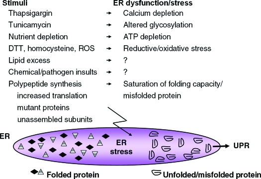
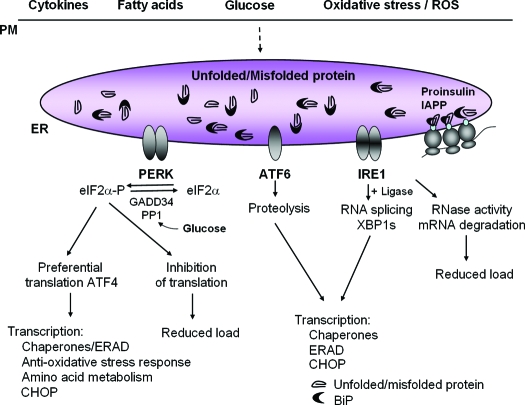
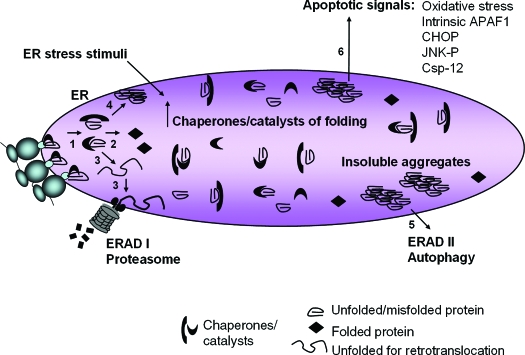
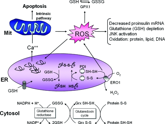
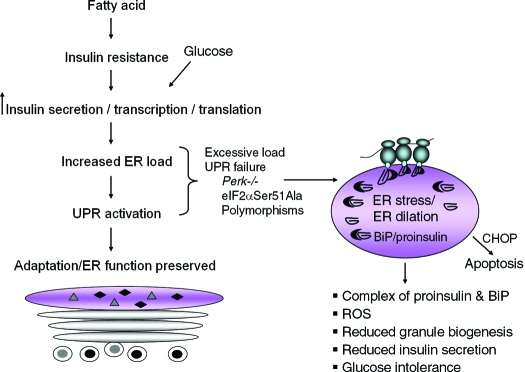
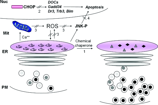
The endoplasmic reticulum (ER) is the entry site into the secretory pathway for newly synthesized proteins destined for the cell surface or released into the extracellular milieu. The study of protein folding and trafficking within the ER is an extremely active area of research that has provided novel insights into many disease processes. Cells have evolved mechanisms to modulate the capacity and quality of the ER protein-folding machinery to prevent the accumulation of unfolded or misfolded proteins. These signaling pathways are collectively termed the unfolded protein response (UPR). The UPR sensors signal a transcriptional response to expand the ER folding capacity, increase degradation of malfolded proteins, and limit the rate of mRNA translation to reduce the client protein load. Recent genetic and biochemical evidence in both humans and mice supports a requirement for the UPR to preserve ER homeostasis and prevent the beta-cell failure that may be fundamental in the etiology of diabetes. Chronic or overwhelming ER stress stimuli associated with metabolic syndrome can disrupt protein folding in the ER, reduce insulin secretion, invoke oxidative stress, and activate cell death pathways. Therapeutic interventions to prevent polypeptide-misfolding, oxidative damage, and/or UPR-induced cell death have the potential to improve beta-cell function and/or survival in the treatment of diabetes.


Figures

Figure 1
Figure 1
Pharmacological and physiological stimuli that cause ER stress and activate the UPR. Thapsigargin, tunicamycin, and dithiothreitol (DTT) are the most common pharmacological agents used to induce ER stress and misfolded protein within the ER lumen in vitro. Physiological stimuli that activate the UPR include nutrient deprivation, elevated lipids or cholesterol, homocysteine (169), numerous chemical insults such as ethanol and nonsteroidal antiinflammatory agents (170,171), viral and bacterial pathogens, and ROS. In addition, increased synthesis of proteins that transit the ER, expression of mutant inherently misfolded proteins, expression of difficult-to-fold polypeptides, or unbalanced expression of subunits of multimeric complexes can cause ER stress, accumulation of unfolded protein, and activation of the UPR. Question marks indicate that the mechanism by which ER stress is generated remains to be elucidated.
Figure 2
Figure 2
The UPR sensors PERK, IRE1α, and ATF6 control mRNA translation and transcriptional induction of UPR-regulated genes. Interaction of BiP with each UPR sensor prevents UPR signaling. Upon accumulation of unfolded protein, BiP is released from each sensor, leading to its activation. The ER protein kinase PERK is activated by homodimerization and autophosphorylation to phosphorylate eIF2α, thereby reducing the rate of mRNA translation and the biosynthetic protein-folding load on the ER. eIF2α phosphorylation paradoxically increases translation of Atf4 mRNA to produce a transcription factor that activates expression of genes encoding protein chaperones, ERAD machinery, enzymes that reduce oxidative stress, and functions in amino acid biosynthesis and transport. Dimerization of the ER protein kinase IRE1α triggers its endoribonuclease activity to induce cleavage of Xbp1 mRNA. Xbp1 mRNA is then ligated by an uncharacterized RNA ligase and translated to produce XBP1s. Concurrently, ATF6 released from BiP transits to the Golgi where it is cleaved to release a transcriptionally active fragment. Cleaved ATF6 acts in concert with XBP1s to induce expression of genes encoding protein chaperones and ERAD machinery. The RNase activity of IRE1α also degrades selective cellular mRNAs to reduce the client protein load upon the ER. Physiological stimuli that can activate the UPR in the β-cell include expression of misfolded proinsulin or IAPP, oxidative stress (ROS), and increases in the extracellular concentrations of glucose, fatty acids, or cytokines.
Figure 3
Figure 3
Pathways of protein misfolding that lead to cell death. Nascent unfolded polypeptides enter the ER and interact with chaperones and catalysts of protein folding to mature into compact, thermodynamically favorable structures, (1) and (2). Failure of this process results in persistence of misfolded polypeptide-chaperone complexes (1) or extraction of soluble, misfolded protein from the ER and degradation through ERAD I (3). Formation of insoluble protein aggregates (4) requires clearance by autophagy (5). ER stress stimuli impair polypeptide folding and induce adaptive increases in chaperones and catalysts within the ER lumen through UPR sensor activation. Chronic or overwhelming stimuli elicit a number of apoptotic signals including oxidative stress, JNK activation, CHOP expression, cleavage of caspase 12, and activation of the intrinsic mitochondrial-dependent cell death pathway (6).
Figure 4
Figure 4
ER stress, protein misfolding, and oxidative stress are intimately interrelated. Protein folding within the ER lumen is ushered by a family of oxidoreductases that catalyze disulfide bond formation and isomerization. ER stress causes an increase in the formation of incorrect intermolecular and/or intramolecular disulfide bonds that require breakage and reformation for proteins to attain the appropriate folded conformation. PDI catalyzes disulfide bond formation and isomerization, whereas glutathione transported into the ER reduces improperly paired disulfide bonds. Reoxidation of PDI is mediated by ERO1; however, ROS are produced in the process. Cellular ROS can deplete glutathione and increase the misfolded protein load in the ER. In turn, ROS can also cause ER stress through modification of proteins and lipids that are necessary to maintain ER homeostasis. Consumption of excessive cellular glutathione due to ER stress could inhibit glutaredoxin reduction and cause accumulation of oxidized cytosolic proteins. ER stress also causes calcium leak from the ER for accumulation in the inner mitochondrial matrix. This calcium loading in the mitochondria can generate additional ROS through disruption of electron transport and opening of the mitochondrial permeability pore. Thus, accumulation of misfolded protein in the ER increases ROS production that can further amplify ER stress, disrupt insulin production, and cause cell death.
Figure 5
Figure 5
UPR signaling preserves ER function and glucose-stimulated insulin secretion (GSIS). Nutrient stimuli and insulin resistance combine to increase the transcription and translation of proinsulin mRNA. The increased biosynthetic load of proinsulin requires UPR signaling for cellular adaptation and maintenance of polypeptide folding within the ER. Excessive biosynthetic load on the ER or genetic defects in UPR signaling, for example Perk-null or eIF2α Ser51Ala mutations, cause ER stress, increase interaction of misfolded proinsulin with BiP, and reduce secretory granule biogenesis. As a consequence, the pool of secretory granules is depleted, and the capacity for GSIS is lost. Humans with polymorphisms that reduce the capacity for protein folding or ER stress signaling may be predisposed to β-cell failure and development of diabetes.
Figure 6
Figure 6
Therapeutic interventions to improve ER function and/or prevent cell death. Potential strategies to improve protein folding in the ER and/or prevent cell death are depicted. Small molecule chemical chaperones may improve polypeptide folding and prevent chronic, irresolvable ER stress (1). As the proapoptotic transcription factor CHOP plays a causal role in ER stress-induced death, strategies to interfere with CHOP induction or function may prevent β-cell failure (2). Because there is a close relationship between oxidative stress and ER stress, antioxidant therapy (3) may increase insulin mRNA expression and translation, as well as improve proinsulin folding to restore GSIS. Finally, JNK activation is a component of the cell death response to both oxidative, and ER stress and inhibition of this death signal may prevent β-cell death (4). It is possible that the reduction of one or more death signals would be sufficient to halt progression to cell death and allow the adaptive functions of the UPR and antioxidant protective mechanisms to preserve productive protein folding within the ER.

References

    1. Ghaemmaghami S, Huh WK, Bower K, Howson RW, Belle A, Dephoure N, O’Shea EK, Weissman JS 2003 Global analysis of protein expression in yeast. Nature 425:737–741 - PubMed
    1. Ma Y, Hendershot LM 2004 ER chaperone functions during normal and stress conditions. J Chem Neuroanat 28:51–65 - PubMed
    1. Kleizen B, Braakman I 2004 Protein folding and quality control in the endoplasmic reticulum. Curr Opin Cell Biol 16:343–349 - PubMed
    1. Ellgaard L 2004 Catalysis of disulphide bond formation in the endoplasmic reticulum. Biochem Soc Trans 32:663–667 - PubMed
    1. Sevier CS, Kaiser CA 2006 Conservation and diversity of the cellular disulfide bond formation pathways. Antioxid Redox Signal 8:797–811 - PubMed

Publication types