. 2008 Aug;149(8):3764-77.
doi: 10.1210/en.2007-1770. Epub 2008 Apr 24.

The role of mineralocorticoid receptor expression in brain remodeling after cerebral ischemia

Affiliations

The role of mineralocorticoid receptor expression in brain remodeling after cerebral ischemia

Naofumi Oyamada et al. Endocrinology. 2008 Aug.

Abstract

Mineralocorticoid receptors (MRs) are classically known to be expressed in the distal collecting duct of the kidney. Recently it was reported that MR is identified in the heart and vasculature. Although MR expression is also found in the brain, it is restricted to the hippocampus and cerebral cortex under normal condition, and the role played by MRs in brain remodeling after cerebral ischemia remains unclear. In the present study, we used the mouse 20-min middle cerebral artery occlusion model to examine the time course of MR expression and activity in the ischemic brain. We found that MR-positive cells remarkably increased in the ischemic striatum, in which MR expression is not observed under normal conditions, during the acute and, especially, subacute phases after stroke and that the majority of MR-expressing cells were astrocytes that migrated to the ischemic core. Treatment with the MR antagonist spironolactone markedly suppressed superoxide production within the infarct area during this period. Quantitative real-time RT-PCR revealed that spironolactone stimulated the expression of neuroprotective or angiogenic factors, such as basic fibroblast growth factor (bFGF) and vascular endothelial growth factor (VEGF), whereas immunohistochemical analysis showed astrocytes to be cells expressing bFGF and VEGF. Thereby the incidence of apoptosis was reduced. The up-regulated bFGF and VEGF expression also appeared to promote endogenous angiogenesis and blood flow within the infarct area and to increase the number of neuroblasts migrating toward the ischemic striatum. By these beneficial effects, the infarct volume was significantly reduced in spironolactone-treated mice. Spironolactone may thus provide therapeutic neuroprotective effects in the ischemic brain after stroke.


Figures
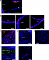
Figure 1
Figure 1
Immunohistochemical examination for MR expression in the brain under normal conditions. A–E, Immunostaining of Neu-N (blue) and MR (red) in the hippocampus (A), CA1, CA2–3 region of the dentate gyrus (B), cerebral cortex (C), and striatum (D and E). Neu-N-positive neurons expressing MR are shown in purple (blue + red). F, Immunostaining of Neu-N (blue), MR (red), and GFAP (green) in the corpus callosum. G, Immunostaining of PECAM-1 (blue), MR (red), and GFAP (green) in the striatum. Scale bar, 500 μm (A and D), magnification, ×5; scale bar, 100 μm (B and C, E and G), magnification, ×20.
Figure 2
Figure 2
Evaluation of the MR-positive area in the ischemic striatum during the acute, subacute, and chronic phases after MCAo. A–E, Representative fluorescence photomicrographs showing immunostaining of Neu-N (blue), MR (red), and GFAP (green) in the ischemic striatum on d 1 (A), 2 (B), 7 (C), 14 (D), and 28 (E) after induction of 20-min MCAo. F and G, Detection of vascular endothelial cells expressing MR by immunostaining PECAM-1 (blue) and MR (red) in the ischemic striatum on d 2 (F) and 7 (G) after MCAo. H, Time-dependent changes in the size of the MR-positive area (square millimeters) in the ischemic striatum. Scale bar, 100 μm (A–G); magnification, ×20.
Figure 3
Figure 3
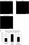
Effects of spironolactone on ROS production in the ischemic striatum on d 7 after 20-min MCAo. A–C, Representative photomicrographs of diHE fluorescence (red), revealing ROS in the nonischemic striatum under normal condition (A) and in the ischemic striatum in vehicle- (B) and spironolactone-treated (C) mice. D, Quantitative analysis of diHE fluorescence intensities in the nonischemic striatum (n = 10) and the ischemic striatum of vehicle- (n = 10) and spironolactone-treated mice (n = 9). ***, P < 0.001. Scale bar, 100 μm (A–C); magnification, ×20.
Figure 4
Figure 4
Antiapoptotic effect of spironolactone on d 7 after 20-min MCAo. A–C, Representative photomicrographs showing immunostaining of ssDNA (green) and Neu-N (blue) in the nonischemic contralateral striatum (A) and in the ischemic ipsilateral striatum in vehicle- (B) and spironolactone-treated (C) mice. D, Quantification of ssDNA-positive apoptotic cells in the nonischemic (n = 10) and ischemic striatum in the vehicle- (n = 11) and spironolactone-treated (n = 10) mice. **, P < 0.01. Scale bar, 100 μm (A–C); magnification, ×20.
Figure 5
Figure 5
Effects of spironolactone on the expression of neuroprotective and angiogenic factors in the ischemic brain on d 2 and 7 after 20-min MCAo. A, Quantitative real-time RT-PCR analysis of BDNF, NGF, GDNF, bFGF, and VEGF levels in the ipsilateral hemisphere in sham-operated (n = 8) and vehicle- (n = 12) and spironolactone-treated (n = 12) mice on d 2 and 7 after MCAo. *, P < 0.05; **, P < 0.01 spironolactone vs. vehicle. B–D, Representative photomicrographs showing immunostaining of bFGF (red), Neu-N (blue), and GFAP (green) on d 7 after MCAo in the nonischemic striatum (B) and the ischemic striatum of vehicle- (C) and spironolactone-treated (D) mice. E–G, Representative photomicrographs showing immunostaining of VEGF (red), Neu-N (blue), and GFAP (green) on d 7 after MCAo in the nonischemic striatum (E) and the ischemic striatum of the vehicle- (F) and spironolactone-treated (G) mice. H, Measurement of the area (square millimeters) of bFGF and VEGF positivity in the nonischemic (n = 3) and ischemic striatum of the vehicle- and spironolactone-treated mice (n = 8–11). **, P < 0.01. Scale bar, 100 μm (B–G); magnification, ×20.
Figure 5
Figure 5
Effects of spironolactone on the expression of neuroprotective and angiogenic factors in the ischemic brain on d 2 and 7 after 20-min MCAo. A, Quantitative real-time RT-PCR analysis of BDNF, NGF, GDNF, bFGF, and VEGF levels in the ipsilateral hemisphere in sham-operated (n = 8) and vehicle- (n = 12) and spironolactone-treated (n = 12) mice on d 2 and 7 after MCAo. *, P < 0.05; **, P < 0.01 spironolactone vs. vehicle. B–D, Representative photomicrographs showing immunostaining of bFGF (red), Neu-N (blue), and GFAP (green) on d 7 after MCAo in the nonischemic striatum (B) and the ischemic striatum of vehicle- (C) and spironolactone-treated (D) mice. E–G, Representative photomicrographs showing immunostaining of VEGF (red), Neu-N (blue), and GFAP (green) on d 7 after MCAo in the nonischemic striatum (E) and the ischemic striatum of the vehicle- (F) and spironolactone-treated (G) mice. H, Measurement of the area (square millimeters) of bFGF and VEGF positivity in the nonischemic (n = 3) and ischemic striatum of the vehicle- and spironolactone-treated mice (n = 8–11). **, P < 0.01. Scale bar, 100 μm (B–G); magnification, ×20.
Figure 6
Figure 6
Effects of spironolactone on vascular regeneration and blood flow in the ischemic striatum after 20-min MCAo. A–E, Histological examination of the vasculature in the ischemic core stained with mouse PECAM-1 (red) and Neu-N (blue). Shown are representative photomicrographs in the nonischemic striatum (A) and the ischemic striatum of vehicle- (B and D) and spironolactone-treated (C and E) mice on d 7 (B and C) and 14 (D and E) after MCAo. F, Quantitative analysis of the relative area of PECAM-1 positivity (percent area) in the nonischemic striatum (n = 5) and ischemic striatum of vehicle- and spironolactone-treated mice (n = 9–10) on d 7 and 14 after MCAo. G–I, Representative photomicrographs of sections of the nonischemic striatum of a sham-operated mouse (G) and the ischemic core of the striatum in vehicle- (H) and spironolactone-treated (I) mice on d 14 after MCAo. J, Quantitative analysis of the relative blood flow in the nonischemic striatum of sham-operated mice (n = 5) and ischemic core of the striatum in vehicle- (n = 11) and spironolactone-treated mice (n = 11) on d 14 after MCAo. The relative blood flow in the ischemic striatum in vehicle- and spironolactone-treated mice was expressed as the ratio of number of fluorescent microspheres in the ischemic core to that in the nonischemic striatum of sham-operated mice (set as 100%). *, P < 0.05 spironolactone vs. vehicle. Scale bar, 100 μm (A–E, G–I); magnification, ×20.
Figure 6
Figure 6
Effects of spironolactone on vascular regeneration and blood flow in the ischemic striatum after 20-min MCAo. A–E, Histological examination of the vasculature in the ischemic core stained with mouse PECAM-1 (red) and Neu-N (blue). Shown are representative photomicrographs in the nonischemic striatum (A) and the ischemic striatum of vehicle- (B and D) and spironolactone-treated (C and E) mice on d 7 (B and C) and 14 (D and E) after MCAo. F, Quantitative analysis of the relative area of PECAM-1 positivity (percent area) in the nonischemic striatum (n = 5) and ischemic striatum of vehicle- and spironolactone-treated mice (n = 9–10) on d 7 and 14 after MCAo. G–I, Representative photomicrographs of sections of the nonischemic striatum of a sham-operated mouse (G) and the ischemic core of the striatum in vehicle- (H) and spironolactone-treated (I) mice on d 14 after MCAo. J, Quantitative analysis of the relative blood flow in the nonischemic striatum of sham-operated mice (n = 5) and ischemic core of the striatum in vehicle- (n = 11) and spironolactone-treated mice (n = 11) on d 14 after MCAo. The relative blood flow in the ischemic striatum in vehicle- and spironolactone-treated mice was expressed as the ratio of number of fluorescent microspheres in the ischemic core to that in the nonischemic striatum of sham-operated mice (set as 100%). *, P < 0.05 spironolactone vs. vehicle. Scale bar, 100 μm (A–E, G–I); magnification, ×20.
Figure 7
Figure 7
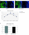
Effects of spironolactone on migration of neuroblasts toward the ischemic striatum after 20-min MCAo. A and B, Immunostaining of Neu-N (blue) and Dcx (red) in the ischemic striatum of vehicle- (A) and spironolactone-treated (B) mice on d 7 after MCAo. C, Quantitative analysis of the numbers (counts per field) of Dcx-positive neuroblasts in the ischemic striatum of vehicle- and spironolactone-treated mice (n = 6/each group). *, P < 0.05. Scale bar, 100 μm (A and B); magnification, ×20.
Figure 8
Figure 8
Effects of spironolactone on infarct size after 20-min MCAo. A and B, Representative fluorescence photomicrographs showing the ischemic striatum of vehicle- (A) and spironolactone-treated (B) mice on d 14 after MCAo. The black area, in which Neu-N-positive neurons are not observed, is the infarcted area. C, Measurement of the infarct areas (square millimeters) in the ischemic striatum in five coronal sections (−1, −0.5, ± 0, +0.5, and +1 mm from bregma) in vehicle- (n = 7) and spironolactone-treated (n = 8) mice. *, P < 0.05; **, P < 0.01 spironolactone vs. vehicle in the corresponding section. D, Measurement of the infarct volume (cubic millimeters) in the two treatment groups. *, P < 0.05. Scale bar, 500 μm (A and B); magnification, ×5.

Comment in

Similar articles

Cited by

References

    1. Sheppard KE, Autelitano DJ 2002 11β-Hydroxysteroid dehydrogenase 1 transforms 11-dehydrocorticosterone into transcriptionally active glucocorticoid in neonatal rat heart. Endocrinology 143:198–204 - PubMed
    1. Takeda Y, Yoneda T, Demura M, Usukura M, Mabuchi H 2002 Calcineurin inhibition attenuates mineralocorticoid-induced cardiac hypertrophy. Circulation 105:677–679 - PubMed
    1. Brilla CG, Matsubara LS, Weber KT 1993 Anti-aldosterone treatment and the prevention of myocardial fibrosis in primary and secondary hyperaldosteronism. J Mol Cell Cardiol 25:563–575 - PubMed
    1. Kuster GM, Kotlyar E, Rude MK, Siwik DA, Liao R, Colucci WS, Sam F 2005 Mineralocorticoid receptor inhibition ameliorates the transition to myocardial failure and decreases oxidative stress and inflammation in mice with chronic pressure overload. Circulation 111:420–427 - PubMed
    1. Virdis A, Neves MF, Amiri F, Viel E, Touyz RM, Schiffrin EL 2002 Spironolactone improves angiotensin-induced vascular changes and oxidative stress. Hypertension 40:504–510 - PubMed

Publication types

MeSH terms