Effects of gestational diethylstilbestrol treatment on male and female gonads during early embryonic development
- PMID: 18436715
- PMCID: PMC2488225
- DOI: 10.1210/en.2007-1599
Effects of gestational diethylstilbestrol treatment on male and female gonads during early embryonic development
Abstract
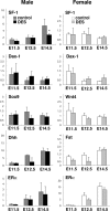
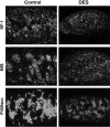
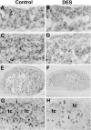
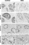
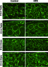
To study the effects of gestational exposure to estrogen on early gonadal differentiation, pregnant mice were treated by sc injection of diethylstilbestrol (DES) or vehicle from embryonic day (E) 8.5 to E14.5, and gonads at E11.5, E12.5, and E14.5 were examined. Quantitative real-time RT-PCR and in situ hybridization revealed that mRNA levels of steroidogenic factor 1 (SF-1), a key regulator of gonadal differentiation, and several male gonad-specific genes, including Müllerian-inhibiting substance (MIS), steroidogenic acute regulatory protein, cholesterol side-chain cleavage cytochrome P450, and Cerebellin 1 precursor protein, were significantly decreased in the DES-treated testis, compared with the control testis at E12.5 and/or E14.5. Immunohistochemistry demonstrated that the staining intensities for SF-1 and MIS in Sertoli cells were apparently reduced in the DES-treated testis, compared with those of the controls, at E12.5 and E14.5. Because MIS, steroidogenic acute regulatory protein, cholesterol side-chain cleavage cytochrome P450, and Cerebellin 1 precursor protein are activated under the regulation of SF-1, the down-regulation of these factors may be due to reduced SF-1 expression. Immunohistochemistry for laminin-1 demonstrated that ovigerous cords in the DES-treated ovary were smaller than those in controls at E14.5. Moreover, the number of 5-bromo-2'deoxyuridine-5-monophosphate-labeled cells in the DES-treated testis was significantly reduced at E12.5 and E14.5, compared with controls, and that in the DES-treated ovary remained higher than that in the control ovary at E14.5. The results suggest that exogenous estrogens can alter sex-specific genetic pathways governing early differentiation and cell proliferation of both male and female gonads.
Figures


Similar articles
-
Exposure of neonatal rats to diethylstilbestrol affects the expression of genes involved in ovarian differentiation.J Med Dent Sci. 2003 Mar;50(1):35-40. J Med Dent Sci. 2003. PMID: 12715917
-
Comparative localization of Dax-1 and Ad4BP/SF-1 during development of the hypothalamic-pituitary-gonadal axis suggests their closely related and distinct functions.Dev Dyn. 2001 Apr;220(4):363-76. doi: 10.1002/dvdy.1116. Dev Dyn. 2001. PMID: 11307169
-
Downregulation of cytochrome P450scc as an initial adverse effect of adult exposure to diethylstilbestrol on testicular steroidogenesis.Environ Toxicol. 2014 Dec;29(12):1452-9. doi: 10.1002/tox.21875. Epub 2013 Jul 20. Environ Toxicol. 2014. PMID: 23873838
-
Developmental expression of mouse steroidogenic factor-1, an essential regulator of the steroid hydroxylases.Mol Endocrinol. 1994 May;8(5):654-62. doi: 10.1210/mend.8.5.8058073. Mol Endocrinol. 1994. PMID: 8058073
-
SF-1: a key regulator of development and function in the mammalian reproductive system.Acta Paediatr Jpn. 1996 Aug;38(4):412-9. doi: 10.1111/j.1442-200x.1996.tb03516.x. Acta Paediatr Jpn. 1996. PMID: 8840555 Review.
Cited by
-
Estrogenic endocrine disruptor exposure directly impacts erectile function.Commun Biol. 2024 Apr 2;7(1):403. doi: 10.1038/s42003-024-06048-1. Commun Biol. 2024. PMID: 38565966 Free PMC article.
-
Effects of low-dose bisphenol AF on mammal testis development via complex mechanisms: alterations are detectable in both infancy and adulthood.Arch Toxicol. 2022 Dec;96(12):3373-3383. doi: 10.1007/s00204-022-03377-0. Epub 2022 Sep 13. Arch Toxicol. 2022. PMID: 36098747
-
Acatalasemic mice are mildly susceptible to adriamycin nephropathy and exhibit increased albuminuria and glomerulosclerosis.BMC Nephrol. 2012 Mar 25;13:14. doi: 10.1186/1471-2369-13-14. BMC Nephrol. 2012. PMID: 22443450 Free PMC article.
-
Effects of in utero exposure to Bisphenol A or diethylstilbestrol on the adult male reproductive system.Birth Defects Res B Dev Reprod Toxicol. 2011 Dec;92(6):526-33. doi: 10.1002/bdrb.20336. Epub 2011 Sep 15. Birth Defects Res B Dev Reprod Toxicol. 2011. PMID: 21922642 Free PMC article.
References
-
- Swain A, Lovell-Badge R 1999 Mammalian sex determination: a molecular drama. Genes Dev 13:755–767 - PubMed
-
- Koopman P, Gubbay J, Vivian N, Goodfellow P, Lovell-Badge R 1991 Male development of chromosomally female mice transgenic for Sry. Nature 351:117–121 - PubMed
-
- Bouma GJ, Hart GT, Washburn LL, Recknagel AK, Eicher EM 2004 Using real time RT-PCR analysis to determine multiple gene expression patterns during XX and XY mouse fetal gonad development. Genes Expr Patterns 5:141–5149 - PubMed
-
- Nef S, Schaad O, Stallings NR, Cederroth CR, Pitetti JL, Schaer G, Malki S, Dubois-Dauphin M, Boizet-Bonhoure B, Descombes P, Parker KL, Vassalli JD 2005 Gene expression during sex determination reveals a robust female genetic program at the onset of ovarian development. Dev Biol 287:361–377 - PubMed
Publication types
MeSH terms
Substances
LinkOut - more resources
Full Text Sources
Molecular Biology Databases

