. 2008 Jun;28(6):1111-6.
doi: 10.1161/ATVBAHA.108.162347. Epub 2008 Apr 24.

Thrombocytopenia and platelet abnormalities in high-density lipoprotein receptor-deficient mice

Affiliations

Thrombocytopenia and platelet abnormalities in high-density lipoprotein receptor-deficient mice

Vandana S Dole et al. Arterioscler Thromb Vasc Biol. 2008 Jun.

Abstract

Objective: High-density lipoprotein (HDL) receptor, scavenger receptor class B, type I (SR-BI), mediated cellular uptake of lipoprotein cholesterol controls HDL structure and plasma HDL and biliary cholesterol levels. In SR-BI knockout (KO) mice, an unusually high plasma unesterified-to-total cholesterol ratio (UC:TC) and abnormally large HDL particles apparently contribute to pathology, including female infertility, susceptibility to atherosclerosis and coronary heart disease, and anemia. Here we examined the influence of SR-BI deficiency on platelets.

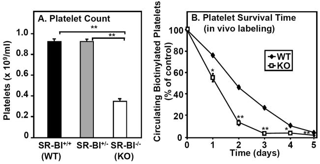
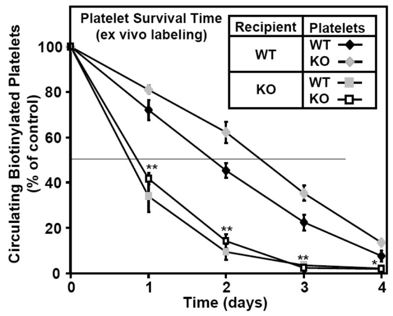
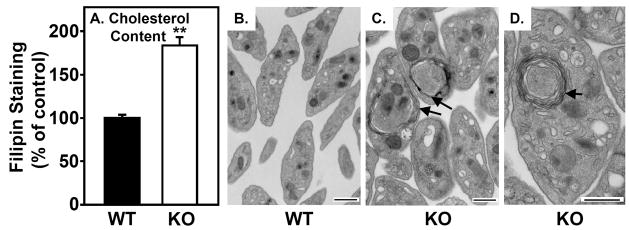
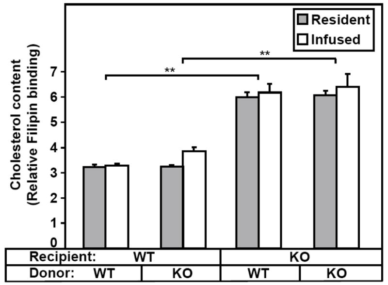
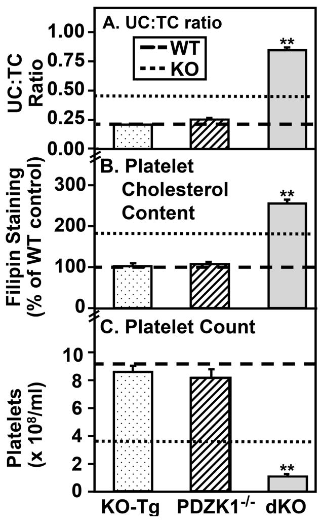
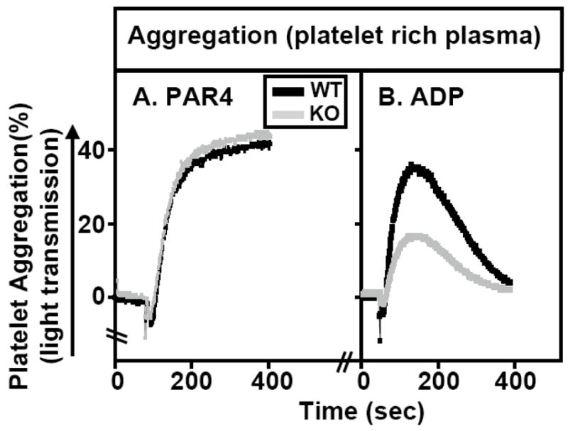
Methods and results: The high plasma UC:TC ratio in SR-BI KO mice was correlated with platelet abnormalities, including high cholesterol content, abnormal morphologies, high clearance rates, and thrombocytopenia. One day after platelets from wild-type mice were infused into SR-BI KO mice, they exhibited abnormally high cholesterol content and clearance rates similar to those of endogenous platelets. Platelets from SR-BI KO mice exhibited in vitro a blunted aggregation response to the agonist ADP but a normal response to PAR4.

Conclusions: In SR-BI KO mice abnormal circulating lipoproteins, particularly their high UC:TC ratio-rather than the absence of SR-BI in platelets themselves-induce defects in platelet structure and clearance, together with a mild defect in function.


Conflict of interest statement

Conflict-of-interest disclosure: None

Figures

Figure 1
Figure 1. Effects of SR-BI deficiency on platelet count and survival
A. Platelets in whole blood harvested from SR-BI+/+ (WT, black bar, n=20), SR-BI+/− (gray bar, n=17) and SR-BI−/− (KO, white bar, n=14) mice were stained (anti-GPIIbIIIa antibody), and counted by flow cytometry. Results are averages from three experiments, each containing a minimum of 3 mice/group; ** P<0.001 B. WT (black diamonds) and SR-BI KO (KO, white squares) mice were intravenously infused with biotin to label platelets in vivo. Mice were then bled each day for five days and the fractions of platelets labeled with biotin were determined by flow cytometry. The percentage of biotinylated platelets was defined as 100% on day 0. (determined 30 minutes after infusion of biotin-NHS) (n=4–5, *P<0.05, **P<0.001).
Figure 2
Figure 2. Survival of ex vivo biotinylated platelets after infusion into WT and KO recipients
Platelets from wild type (WT, black diamonds or gray squares) or SR-BI KO (white squares or gray diamonds) mice were isolated, washed, biotinylated and intravenously infused into recipient mice of the indicated phenotypes (WT, diamonds; SR-BI KO, squares), and the percentage of labeled platelets in the circulation relative to day 0 immediately after infusion (100% of control) was determined as in Figure 1B. Results represent the average of two independent experiments (n=6–7). Values for donors of either genotype infused into SR-BI KO recipients were significantly lower than for WT recipients (*P<0.05 and **P<0.001).
Figure 3
Figure 3. Cholesterol content and ultrastructure of platelets
A. Platelets in blood from WT (black bar) and SR-BI KO (white bar) mice were stained with anti-GPIIbIIIa, then incubated with 50 μg/ml filipin (15 minutes, RT) to label unesterified cholesterol, and filipin staining was determined by flow cytometry (n=6–7, **P<0.001). B–D. Washed platelets from WT (B) and SR-BI KO (C, D) mice were visualized using standard transmission electron microscopy. Multi-lamellar, membrane-like structures often seen in SR-BI KO platelets are indicated by arrows. Scale bar = 500 nm.
Figure 4
Figure 4. Cholesterol content of ex vivo biotinylated platelets after infusion into WT or SR-BI KO recipients
Platelets from WT or SR-BI KO mice were labeled with biotin and infused into WT or SR-BI KO recipients. Next day the recipient mice were bled, the platelets stained with GPIIbIIIa-APC, PE-SA and filipin, and the cholesterol content (filipin staining) of resident (non-biotinylated) and infused platelets was determined by flow cytometry. (n=3–4). The data are from one of two similar independent experiments (**P<0.001).
Figure 5
Figure 5. Effects of altering SR-BI, PDZK1 and apoE expression on plasma UC:TC ratio, platelet cholesterol content and platelet count
Blood was drawn from WT mice (dashed lines), SR-BI KO mice (dotted lines), SR-BI KO mice expressing a primarily liver specific SR-BI transgene (KO-Tg, stippled bars), PDZK1−/− mice (hatched bars), and SR-BI KO/apoE−/− double knockout mice (dKO, gray bars) and plasma unesterified-to-total cholesterol ratios (UC:TC) (panel A, n=7–18), platelet cholesterol contents (panel B, filipin staining, n=7) and platelet counts (panel C, n=7–13) were determined (**P<0.001).
Figure 6
Figure 6. Effects of platelet agonists PAR4 peptide and ADP on platelet aggregation
Platelet-rich plasma from WT (black lines) and SR-BI KO (gray lines) were isolated and platelet aggregation as a function of time after adding the agonists PAR4 peptide (1 mM, panel A) or ADP (1 μM, panel B) was measured as increased light transmission in an aggregometer as described in Methods. Aggregation traces from one of three independent experiments are shown.

Similar articles

Cited by

References

    1. Acton S, Rigotti A, Landschulz KT, Xu S, Hobbs HH, Krieger M. Identification of scavenger receptor SR-BI as a high density lipoprotein receptor. Science. 1996;271:518–520. - PubMed
    1. Rigotti A, Miettinen HE, Krieger M. The role of the high-density lipoprotein receptor SR-BI in the lipid metabolism of endocrine and other tissues. Endocr Rev. 2003;24:357–387. - PubMed
    1. Zannis VI, Chroni A, Krieger M. Role of apoA-I, ABCA1, LCAT, and SR-BI in the biogenesis of HDL. J Mol Med. 2006;84:276–294. - PubMed
    1. Kocher O, Yesilaltay A, Cirovic C, Pal R, Rigotti A, Krieger M. Targeted disruption of the PDZK1 gene in mice causes tissue-specific depletion of the high density lipoprotein receptor scavenger receptor class B type I and altered lipoprotein metabolism. J Biol Chem. 2003;278:52820–52825. - PubMed
    1. Rigotti A, Trigatti BL, Penman M, Rayburn H, Herz J, Krieger M. A targeted mutation in the murine gene encoding the high density lipoprotein (HDL) receptor scavenger receptor class B type I reveals its key role in HDL metabolism. Proc Natl Acad Sci U S A. 1997;94:12610–12615. - PMC - PubMed

Publication types

MeSH terms