. 2008 Oct;26(10):2682-90.
doi: 10.1634/stemcells.2008-0030. Epub 2008 Apr 24.

IFATS collection: The role of human adipose-derived stromal cells in inflammatory microvascular remodeling and evidence of a perivascular phenotype

Affiliations

IFATS collection: The role of human adipose-derived stromal cells in inflammatory microvascular remodeling and evidence of a perivascular phenotype

Peter J Amos et al. Stem Cells. 2008 Oct.

Abstract

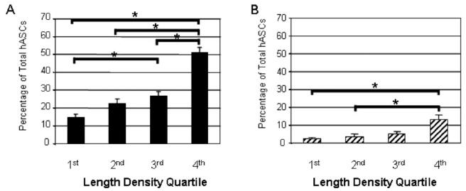
A growing body of literature suggests that human adipose-derived stromal cells (hASCs) possess developmental plasticity both in vitro and in vivo, and might represent a viable cell source for therapeutic angiogenesis and tissue engineering. We investigate their phenotypic similarity to perivascular cell types, ability to contribute to in vivo microvascular remodeling, and ability to modulate vascular stability. We evaluated hASC surface expression of vascular and stem/progenitor cell markers in vitro, as well as any effects of platelet-derived growth factor B chain (PDGF-BB) and vascular endothelial growth factor 165 on in vitro hASC migration. To ascertain in vivo behavior of hASCs in an angiogenic environment, hASCs were isolated, expanded in culture, labeled with a fluorescent marker, and injected into adult nude rat mesenteries that were stimulated to undergo microvascular remodeling. Ten, 30, and 60 days after injection, tissues from anesthetized animals were harvested and processed with immunohistochemical techniques to determine hASC quantity, positional fate in relation to microvessels, and expression of endothelial and perivascular cell markers. After 60 days, 29% of hASCs exhibited perivascular morphologies compared with 11% of injected human lung fibroblasts. hASCs exhibiting perivascular morphologies also expressed markers characteristic of vascular pericytes: smooth muscle alpha-actin (10%) and neuron-glia antigen 2 (8%). In tissues treated with hASCs, vascular density was significantly increased over age-matched controls lacking hASCs. This study demonstrates that hASCs express pericyte lineage markers in vivo and in vitro, exhibit increased migration in response to PDGF-BB in vitro, exhibit perivascular morphology when injected in vivo, and contribute to increases in microvascular density during angiogenesis by migrating toward vessels. Disclosure of potential conflicts of interest is found at the end of this article.


Figures

Figure 1
Figure 1. In Vitro Characterization of hASC Surface Marker Expression
(A) Average expression (± standard deviation) of CD31 (white) and CD144 (black) is minimal and declines to 0%. (B) Detected expression of CD34 depends on the specific antibody used (8G12: white, 581: black, BI-3C5: gray). (C) Average expression (± standard deviation) of NG2 using flow cytometry.
Figure 2
Figure 2. In Vitro Migration of hASCs in the Presence of PDGF-BB or VEGF165
(A) Relative hASC migration (± standard deviation; * = p<0.001) into untreated scratch wounds or wounds treated with VEGF165 or PDGF-BB. (B) Inhibition of PDGF-BB-induced migration using antibodies to PDGF-BB and PDGF receptor β (± standard deviation; * = p<0.001; ‡ = p<0.005; † = p<0.01; # = p<0.05).
Figure 3
Figure 3. Pericyte-like Localization and Surface Marker Expression of hASCs in Rat Mesentery
Rat mesenteric tissues injected with human cells were harvested 60 days later and immunostained to visualize microvessels. A) Some DiI(+) hASCs express NG2 (yellow, arrows), some do not (red), and some exhibit pericyte-like morphologies (vessel wrapping) and are aligned with capillaries that express BSI-lectin (blue). B) Some hASCs expressing SMA (yellow) and exhibiting pericyte-like morphologies (arrows) along BSI-lectin-positive capillaries (blue). C-D) Examples of DiI-labeled SMA-expressing hASCs (yellow) in mesenteric tissue co-stained with SMA and BSI-lectin (both stains are green). E) Some hLFs (red) exhibit pericyte-like morphologies (arrows) along BSI-lectin-positive capillary (blue) and SMA-positive venules (green), although hLFs do not express SMA and total vessel density is reduced compared to hASC-injected tissue, as seen in B. F) No DiI(+) cells are present in control, un-injected tissues (green: BSI-lectin). Scale bar = 25 μm in A,B,E,F; scale bar = 20 μm in C,D.
Figure 4
Figure 4. Quantification of In Vivo Pericyte-like Localization and Surface Marker Expression in 48-80-stimulated tissues
(A) Percentage of hASCs and hLFs that exhibit pericyte-like morphologies regardless of pericyte marker expression. (B) Percentage of hASCs and hLFs in the tissue that exhibit pericyte-like morphologies (but not pericyte markers). (C) Percentage of total hASCs in the tissue that express pericyte markers (but not morphology). (D) Percentage of total hASCs in the tissue that both exhibit pericyte-like morphologies and express pericyte markers. Error bars represent standard error; * = (p≤0.05).
Figure 5
Figure 5. Functional Impact of hASCs on Vascular Length Density In Vivo
Vascular length density for vehicle control (no cells: white) vs. hASC (black) vs. hLF (gray) in (A) 48-80-treated tissues and (B) tissues not treated with 48-80. Error bars represent standard error; * = p≤0.05.
Figure 6
Figure 6. Distribution of hASCs With Respect to Length Density Quartile
A) Percentage of total hASCs in the tissue that exhibit pericyte-like-morphologies at day 60, and B) percentage of total hASCs that exhibit pericyte-like morphologies and also express pericyte markers for each quartile of length density at day 60. Error bars represent standard error; * = significantly different (p≤0.05).

References

    1. Iba O, Matsubara H, Nozawa Y, et al. Angiogenesis by implantation of peripheral blood mononuclear cells and platelets into ischemic limbs. Circulation. 2002;106:2019–2025. - PubMed
    1. Peichev M, Naiyer AJ, Zhu Z, et al. Expression of VEGFR-2 and AC133 by circulating human CD34+ cells identifies a population of functional endothelial precursors. Hemostas, Thombosis, and Vasc Biol. 2000;95:952–958. - PubMed
    1. Asahara T, Haruchika M, Tomono T, et al. Bone marrow origin of endothelial progenitor cells responsible for postnatal vasculogenesis in physiological and pathological neovascularization. Circ Res. 1999;85:221–228. - PubMed
    1. Rajantie I, Ilmonen M, Alminaite A, et al. Adult bone marrow-derived cells recruited during angiogenesis comprise precursors for periendothelial vascular mural cells. Blood. 2004;104:2084–2086. - PMC - PubMed
    1. Carmeliet P, Luttun A. The role of the bone marrow-derived stem cells in (therapeutic) angiogenesis. Thrombos & Haemostas. 2001;86:289–297. - PubMed

Publication types

MeSH terms