Review
. 2008 Jun;129(6):667-86.
doi: 10.1007/s00418-008-0427-6. Epub 2008 Apr 24.

The mammalian centrosome and its functional significance

Affiliations
Review

The mammalian centrosome and its functional significance

Heide Schatten. Histochem Cell Biol. 2008 Jun.

Abstract

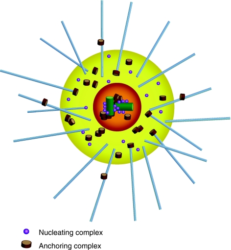
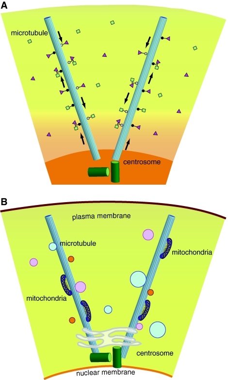
Primarily known for its role as major microtubule organizing center, the centrosome is increasingly being recognized for its functional significance in key cell cycle regulating events. We are now at the beginning of understanding the centrosome's functional complexities and its major impact on directing complex interactions and signal transduction cascades important for cell cycle regulation. The centrosome orchestrates entry into mitosis, anaphase onset, cytokinesis, G1/S transition, and monitors DNA damage. Recently, the centrosome has also been recognized as major docking station where regulatory complexes accumulate including kinases and phosphatases as well as numerous other cell cycle regulators that utilize the centrosome as platform to coordinate multiple cell cycle-specific functions. Vesicles that are translocated along microtubules to and away from centrosomes may also carry enzymes or substrates that use centrosomes as main docking station. The centrosome's role in various diseases has been recognized and a wealth of data has been accumulated linking dysfunctional centrosomes to cancer, Alstrom syndrome, various neurological disorders, and others. Centrosome abnormalities and dysfunctions have been associated with several types of infertility. The present review highlights the centrosome's significant roles in cell cycle events in somatic and reproductive cells and discusses centrosome abnormalities and implications in disease.


Figures

Fig. 1
Fig. 1
A typical mammalian centrosome is composed of two centrioles surrounded by a meshwork of a proteins embedded in matrix called the pericentriolar material (PCM). Gamma-tubulin and the gamma-tubulin ring complex that nucleate microtubules along with associated proteins are embedded in the PCM. Highlighted in this diagram are two centrosomal complexes, the microtubule nucleating complex and the microtubule anchoring complex
Fig. 2
Fig. 2
a The centrosome serves as central station to mediate substrate and enzyme activities along microtubules towards the minus ends of microtubules driven by dynein (white circles) and toward the plus ends driven by kinesin (black circles). b Through its microtubule-organizing capabilities the centrosome mediates distribution of cell organelles such as mitochondria, Golgi, and vesicles of various sizes, many containing regulatory enzymes for cell cycle regulation
Fig. 3
Fig. 3
The structural cell cycle-dependent changes of centrosomes are shown here in sea urchin eggs during the first cell cycle after fertilization. This system had been used by Theodor Boveri for the majority of his classic studies on centrosomes. Centrosome material disperses around the zygote nucleus (aarrows) and separates to the poles during prophase (carrows). Centrosomes become densely compacted in metaphase (earrows) and disperse during anaphase (garrows). The correlated images for microtubules either from the same cell or from corresponding cell cycle stages are shown in b, d, f, and h. Immunofluorescence microscopy of centrosomes, microtubules, and DNA (blue). Centrosomes are displayed in green; microtubules are displayed in green (b, d) or red (f, h). Reprinted with permission from Schatten et al.
Fig. 4
Fig. 4
In interphase, a single centrosome is juxta-positioned to the nucleus. a, c, and d show small GFP-centrin-labeled centrosomes in mouse 3T3 cells. Microtubules are detected with α-tubulin and shown in red; b is of an LNCaP prostate cancer cell labeled with human autoimmune antibody SPJ displaying multiple centrosomal foci perhaps indicating centrosome abnormalities; e shows a porcine fibroblast cell labeled with γ-tubulin to detect the centrosome and Mitotracker Rosamine to detect mitochondria. Microtubules are shown in green; b reprinted with permission from Schatten et al.
Fig. 5
Fig. 5
A typical mammalian centrosome cycle within the cell cycle. The single interphase centrosome (a) is closely associated with the nucleus and nucleates an array of interphase microtubules. Centrosome duplication occurs during the S phase in synchrony with DNA dupliction (b). Centrosome separation of the duplicated centrosome toward opposite poles takes place in the early prophase stage (c). The bipolar mitotic apparatus becomes established when centrosomes have reached the opposite poles and the nuclear envelope has broken down (d). During this stage interphase centrosomes mature into mitotic centrosomes acquiring mitosis-associated centrosomal proteins including NuMA that moves out of the nucleus to become a mitotic centrosome-associated protein. The metaphase centrosome (e) becomes highly compacted to organize the metaphase spindle with microtubules attached to the kinetochores. Anaphase is the stage when centrosomal material becomes decompacted again (f) before reorganizing into interphase centrosomes that associate with the nuclei of the separating daughter cells (g). Centrosomes shaded yellow with centrioles and PCM displayed in black; nuclei shaded orange; microtubules displayed as black rods; chromosomes displayed in black. Modified from Sun and Schatten
Fig. 6
Fig. 6
The chaotropic agent formamide causes centrosomal damage and results in tripolar (a) or multiple (b) centrosomal foci as shown here in an example using sea urchin eggs as model system. Centrosomes labeled red (a) or green (b). Microtubules in b labeled red a= modified from Schatten et al.
Fig. 7
Fig. 7
A tetrapolar spindle displaying microtubules (green) and chromosomes (red) as a result of dispermy in a sea urchin egg. Such results on dispermic eggs had stimulated Boveri to propose that centrosome abnormalities are at the core of malignant tumors. Image produced in collaboration at the Integrated Microscopy Resource at the University of Wisconsin

References

    1. {'text': '', 'ref_index': 1, 'ids': [{'type': 'PMC', 'value': 'PMC2133110', 'is_inner': False, 'url': 'https://pmc.ncbi.nlm.nih.gov/articles/PMC2133110/'}, {'type': 'PubMed', 'value': '10209026', 'is_inner': True, 'url': 'https://pubmed.ncbi.nlm.nih.gov/10209026/'}]}
    2. Ahmad FJ, Yu W, McNally FJ, Baas PW (1999) An essential role for katanin in severing microtubules in the neuron. J Cell Biol 145:305–315 - PMC - PubMed
    1. {'text': '', 'ref_index': 1, 'ids': [{'type': 'PubMed', 'value': '14654843', 'is_inner': True, 'url': 'https://pubmed.ncbi.nlm.nih.gov/14654843/'}]}
    2. Andersen JS, Wilkinson CJ, Mayor T, Mortensen P, Nigg EA, Mann M (2003) Proteomic characterization of the human centrosome by protein correlation profiling. Nature 426:570–574 - PubMed
    1. {'text': '', 'ref_index': 1, 'ids': [{'type': 'PubMed', 'value': '9704896', 'is_inner': True, 'url': 'https://pubmed.ncbi.nlm.nih.gov/9704896/'}]}
    2. Baas PW (1998) The role of motor proteins in establishing the microtubule arrays of axons and dendrites. J Chem Neuroanat 14:175–180 - PubMed
    1. {'text': '', 'ref_index': 1, 'ids': [{'type': 'PubMed', 'value': '10027286', 'is_inner': True, 'url': 'https://pubmed.ncbi.nlm.nih.gov/10027286/'}]}
    2. Baas PW (1999) Microtubules and neuronal polarity: lessons from mitosis. Neuron 22:23–31 - PubMed
    1. {'text': '', 'ref_index': 1, 'ids': [{'type': 'PubMed', 'value': '15738963', 'is_inner': True, 'url': 'https://pubmed.ncbi.nlm.nih.gov/15738963/'}]}
    2. Badano JL, Teslovich TM, Katsanis N (2005) The centrosome in human genetic disease. Nat Rev Genet 6:194–205 - PubMed

Substances

LinkOut - more resources