Review
. 2008 Jun;13(2):235-46.
doi: 10.1007/s10911-008-9080-x. Epub 2008 Apr 25.

ErbB4/HER4: role in mammary gland development, differentiation and growth inhibition

Affiliations
Review

ErbB4/HER4: role in mammary gland development, differentiation and growth inhibition

Rebecca S Muraoka-Cook et al. J Mammary Gland Biol Neoplasia. 2008 Jun.

Abstract

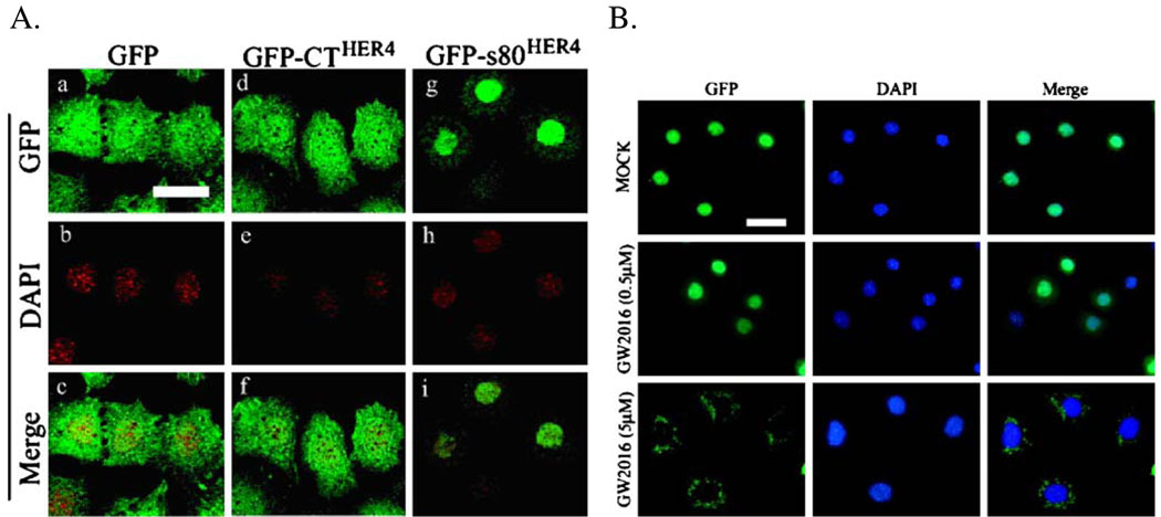
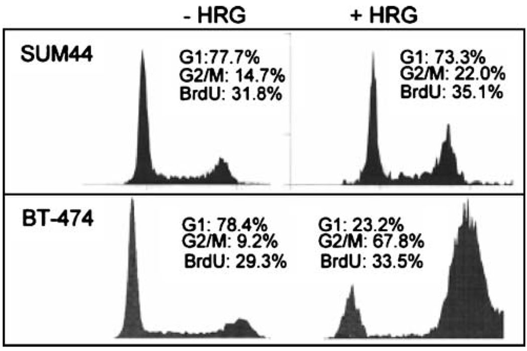
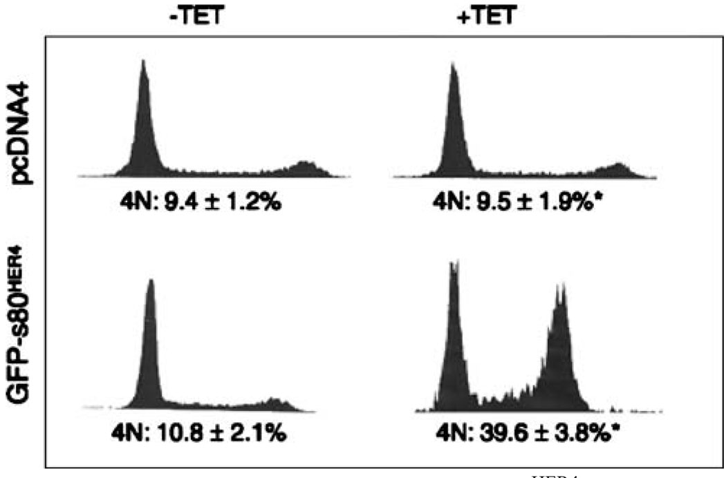
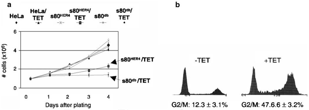
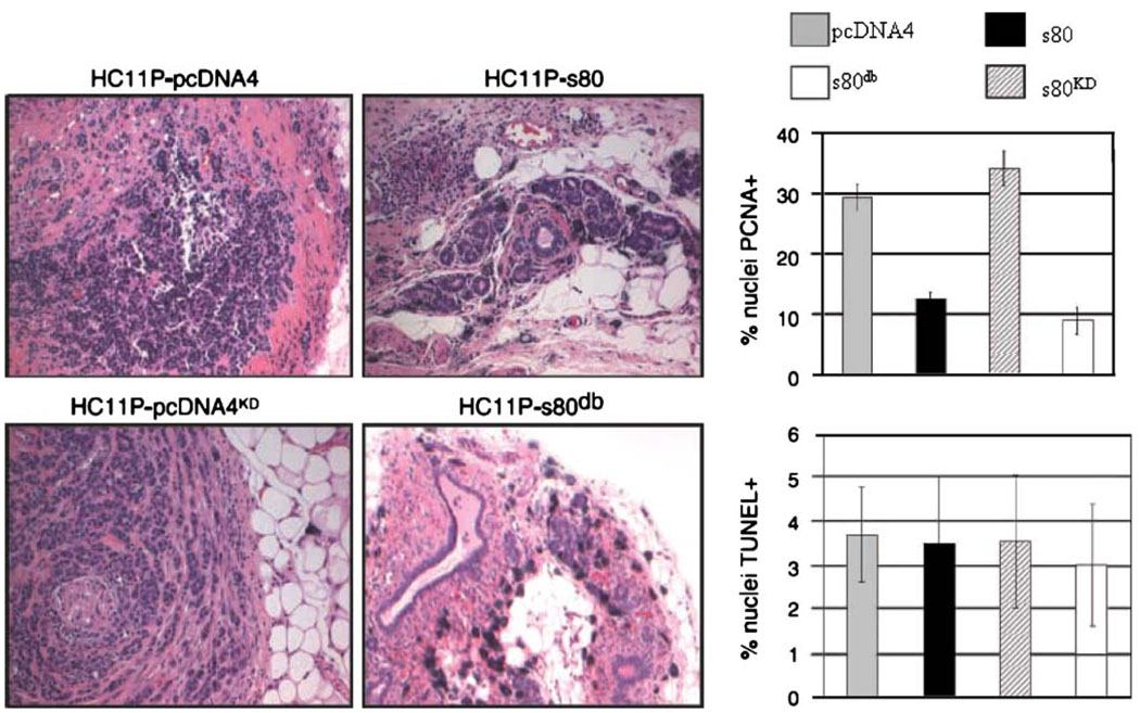
The ErbB receptor tyrosine kinase family has often been associated with increased growth of breast epithelial cells, as well as malignant transformation and progression. In contrast, ErbB4/HER4 exhibits unique attributes from a two step proteolytic cleavage which releases an 80 kilodalton, nuclear localizing, tyrosine kinase to a signal transduction mechanism that slows growth and stimulates differentiation of breast cells. This review provides an overview of ErbB4/HER4 in growth and differentiation of the mammary epithelium, including its physiologic role in development, the contrasting growth inhibition/tumor suppression and growth acceleration of distinct ErbB4/HER4 isoforms and a description of the unique cell cycle regulated pattern of nuclear HER4 ubiquitination and destruction.


Figures

Figure 1
Figure 1
Nuclear localization of HER4/ErbB4 in mouse HC11 cells. a Localization of GFP fluorescence in HC11 cells stably expressing GFP, GFP-CTHER4 (the C-terminal residues 989–1308 of HER4/ErbB4, lacking the kinase domain), or GFP-s80HER4. b HC11 cells stably expressing GFP-s80HER4 were mock treated or treated for 24 h with 0.5 µM GW572016 (a dose that does not abolish s80HER4 tyrosine kinase activity) or 5 µM GW572016 (a dose sufficient to substantially reduce s80HER4 tyrosine kinase activity). The counter-stain with DAPI is shown as is the merge of the two images (GFP and DAPI). Bar=30 µm, for both (a) and (b).
Figure 2
Figure 2
Heregulin-mediated growth inhibition of HER4 positive breast cancer cells. Representative histograms of human SUM44 or BT-474 cells cultured in serum-free medium with or without HRG for 72 h then stained with propidium iodide. In addition, cells were labeled with BrdU for 6 h, stained with an anti-BrdU antibody, and analyzed by flow cytometry. The percentages of the cell population in G1, G2/M phases of the cell cycle and BrdU-positive cells are shown.
Figure 3
Figure 3
The intracellular domain of HER4, s80HER4, is expressed from a tetracycline inducible promoter in HeLa cells. Induction of s80HER4 inhibits growth. Representative histograms of HeLa-pcDNA4 and HeLa-s80Her4 cells grown in serum-free medium ± tetracycline (48 h) and stained with propidium iodide and analyzed by flow cytometry. Percentage of cells in the G2/M phase of the cell cycle are shown, *P<0.002, Student’s unpaired t test.
Figure 4
Figure 4
Mutation of the s80HER4 D-box enhances s80HER4-mediated growth inhibition in tetracycline inducible HeLa cells. a Equal numbers of cells were plated at day 0 ± tetracycline. Cells were counted at 1-day intervals through day 4. P=0.011, comparing cell number of HeLa-s80HER4 + tetracycline at day 4 with cell number of HeLa-s80db + tetracycline at day 4, n=5, analyzed in triplicate. b Analysis of HeLa-s80db cells cultured ± tetracycline (48 h), stained with propidium iodide. Percent of cells in the G2/M phase of the cell cycle are shown.
Figure 5
Figure 5
Decreased tumor formation in PyVmT-transformed HC11 cells (HC11P) expressing s80db. a HC11P cells expressing s80HER4, s80KD (kinase dead), s80db (D-Box mutant), or the empty pcDNA4 vector were injected into the inguinal mammary fat pads of female BALB/c mice. Fifteen weeks after implantation of cells, mice were euthanized and the mammary glands were analyzed histologically. b Five tumor samples per group were analyzed by immunohistochemistry. PCNA-positive (top) or TUNEL-positive (bottom) nuclei. HC11P-s80db percentage of PCNA-positive cells was the lowest of all groups (9.0±2.2) and was also significantly decreased when compared with HC11P-s80 (P<0.037).

References

    1. Cohen S. Isolation of a mouse submaxillary gland protein accelerating incisor eruption and eyelid opening in the new-born animal. J Biol Chem USA. 1962;237:1555–1562. - PubMed
    1. Downward J, Yarden Y, Mayes E, Scrace G, Totty N, Stockwell P, et al. Close similarity of epidermal growth factor receptor and v-erb-B oncogene protein sequences. Nature. 1984;307:521–527. - PubMed
    1. Carpenter G, King L, Jr, Cohen S. Rapid enhancement of protein phosphorylation in A-431 cell membrane preparations by epidermal growth factor. J Biol Chem. 1979;254:4884–4891. - PubMed
    1. Earp HS, Dawson TL, Li X, Yu H. Heterodimerization and functional interaction between EGF receptor family members: a new signaling paradigm with implications for breast cancer research. Breast Cancer Res Treat. 1995;35:115–132. - PubMed
    1. Yarden Y, Sliwkowski MX. Untangling the ErbB signaling network. Nat Rev Mol Cell Biol. 2001;2:127–137. - PubMed

MeSH terms