. 2008 May;15(5):523-30.
doi: 10.1038/nsmb.1417. Epub 2008 Apr 27.

Mouse Eri1 interacts with the ribosome and catalyzes 5.8S rRNA processing

Affiliations

Mouse Eri1 interacts with the ribosome and catalyzes 5.8S rRNA processing

K Mark Ansel et al. Nat Struct Mol Biol. 2008 May.

Abstract

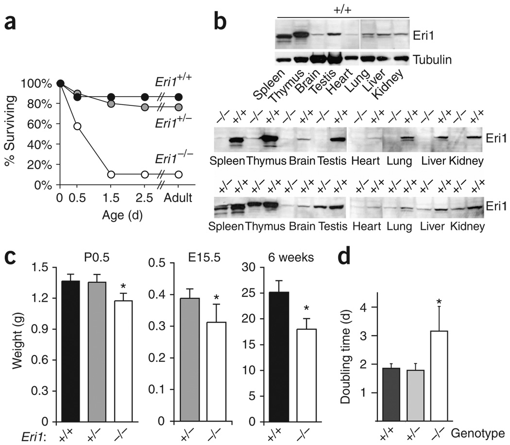
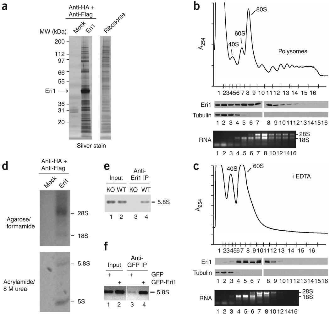
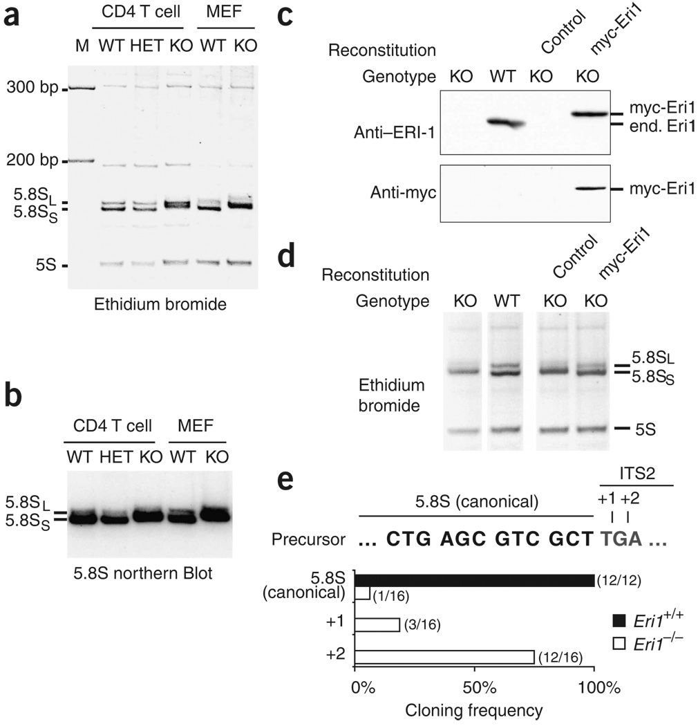
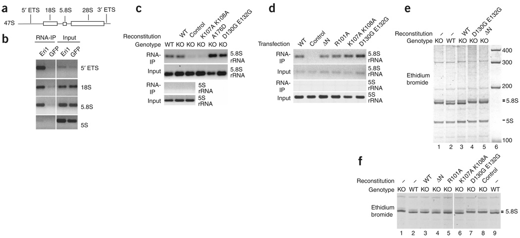
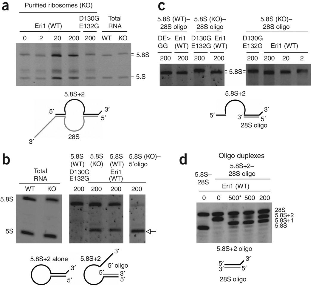
Eri1 is a 3'-to-5' exoribonuclease conserved from fission yeast to humans. Here we show that Eri1 associates with ribosomes and ribosomal RNA (rRNA). Ribosomes from Eri1-deficient mice contain 5.8S rRNA that is aberrantly extended at its 3' end, and Eri1, but not a catalytically inactive mutant, converts this abnormal 5.8S rRNA to the wild-type form in vitro and in cells. In human and murine cells, Eri1 localizes to the cytoplasm and nucleus, with enrichment in the nucleolus, the site of preribosome biogenesis. RNA binding residues in the Eri1 SAP and linker domains promote stable association with rRNA and thereby facilitate 5.8S rRNA 3' end processing. Taken together, our findings indicate that Eri1 catalyzes the final trimming step in 5.8S rRNA processing, functionally and spatially connecting this regulator of RNAi with the basal translation machinery.


Figures

Figure 1
Figure 1
Reduced survival and weight of Eri1-deficient mice. (a) Survival curve of C57BL/6 Eri1−/− mice (open circles), Eri1+/− (gray circles) and wild-type (black circles) littermates. (b) Monoclonal anti-Eri1 immunoblot of NMRI × C57BL/6 F2 Eri1−/−, Eri1+/+ and Eri1+/− mouse tissue extracts. (c) Reduced weight (mean ± s.d.) of C57BL/6 Eri1−/− mice (white), Eri1+/− (gray) and wild-type (black) control littermates on the first morning after birth (P0.5), embryonic day 15.5 (E15.5), and 6 weeks after birth. *, significantly lower weight (one-tailed Student’s t-test, P-values 8 × 10⁻⁷ (P0.5), 0.001 (E15.5), 0.0004 (6 weeks)). (d) Reduced growth of primary Eri1−/− MEFs. Mean (± s.d.) doubling times for three independent experiments using two measurements obtained between passages 6 and 9 of fibroblasts derived from a total of six Eri1−/− embryos and controls. *, significantly increased doubling time (ANOVA P = 0.006).
Figure 2
Figure 2
Eri1 associates with ribosomes. (a) Silver-stained proteins copurified in Eri1 TAP from cytoplasmic lysates of HeLa cells stably transduced with HA-Flag–Eri1 or untransduced cells (Mock). Ribosomes prepared from untransduced HeLa cells were run on the same gel. MW, molecular weight. (b,c) Sucrose gradient fractionation of 3T3 lysates prepared in the absence (b) or presence (c) of 20 mM EDTA. Above, absorbance profiles at 254 nm (A254) obtained while fractionating sucrose gradients from top to bottom. Approximate positions of collected fractions are indicated on the x axis. Middle, polyclonal anti-Eri1 and anti–α-tubulin immunoblots. Below, RNA from the indicated fractions. Protein fractions were run on two gels per gradient, and immunoblots for all four gels were performed in unison. (d) RNA copurified in Eri1 TAP as in a separated by agarose (above) and polyacrylamide (below) gel electrophoresis. (e,f) RNA immunoprecipitation (RIP) assays for Eri1 binding to 5.8S rRNA. (e) RIP with anti-Eri1 monoclonal antibody in Eri1−/− (KO) and wild-type (WT) MEFs. (f) RIP with anti-GFP polyclonal antibody in HEK 293 cells transduced with LNCX2-GFP or LNCX2-GFP–Eri1.
Figure 3
Figure 3
Eri1 is required for 5.8S rRNA 3′ end processing. (a,b) RNA from Eri1+/+ CD4-Cre (WT), Eri1+/fl CD4-Cre (HET) and Eri1fl/fl CD4-Cre (KO) primary CD4+ T cells, and Eri1+/+ (WT) and Eri1−/− (KO) MEFs, visualized by ethidium bromide staining (a) and northern blot for 5.8S rRNA (b). (c) Polyclonal anti-Eri1 and monoclonal anti-myc immunoblot analysis of MEFs of the indicated genotypes left untransduced or transduced with KMV (control) or KMV–myc-Eri1. end., endogenous. (d) Ethidium bromide–stained RNA from the same MEF cell populations. All lanes were run on the same gel. (e) Sequences of cloned 5.8S rRNA 3′ ends. Histogram shows the frequency of sequences obtained from Eri1+/+ (black bars; n = 12) and Eri1−/− (white bars; n = 16) that correspond to the predicted 5.8S sequence or extended sequences as shown above. All sequence extensions matched the expected ITS2 sequences (+1, T; +2, TG), except for one +2 clone that contained a TT extension.
Figure 4
Figure 4
Eri1 binds directly to rRNA and rRNA precursors, and Eri1 exonuclease activity is required for 5.8S rRNA 3′ end processing. (a) Simplified schematic depiction of the 47S rRNA precursor. ETS, external transcribed spacer. (b) RIP assay performed with anti-GFP polyclonal antibody and HEK 293 cells stably transduced with LNCX2-GFP–Eri1 (Eri1) or LNCX2-GFP (GFP). Primers specific for the 5′ ETS, 18S and 5.8S rRNA were used to identify Eri1 association with the 45S rRNA precursor. (c) RIP assay for Eri1 binding to 5.8S rRNA performed with anti-Eri1 monoclonal antibody and wild-type (WT) and Eri1−/− (KO) MEFs left untransduced or reconstituted by transduction with MSCV encoding myc-Eri1 (WT) and the indicated mutants (Supplementary Fig. 7a). (d) RIP assay for overexpressed GFP-Eri1 binding to 5.8S rRNA using anti-GFP polyclonal antibody and HEK 293 cells transiently transfected with pDEST12.2 expression constructs for GFP-Eri1 (WT), the indicated mutants or GFP-expressing vector (control). (e) RNA from WT and KO MEFs left untransduced (−) or reconstituted with MSCV retroviruses encoding myc-Eri1 (WT) and the indicated mutants. (f) RNA from WT and KO MEFs left untransduced (−) or reconstituted with MSCV retroviruses encoding GFP-Eri1 (WT), the indicated mutants, or GFP alone (control). GFP-expressing cells were FACS sorted for equal GFP fluorescence 1 week before RNA extraction (Supplementary Fig. 7c,d). All lanes were run on the same gel.
Figure 5
Figure 5
Eri1 catalyzes 3′ end processing of 5.8S rRNA. RNA was subjected to 30 min in vitro processing reactions with the indicated amounts (ng) of recombinant Eri1 or catalytically inactive Eri1 (D130G E132G). Diagrams depict the predicted secondary structure of unprocessed substrates. (a) Ribosomes purified from Eri1−/− liver. Total RNA from wild-type (WT) and Eri1−/− (KO) testis was included to show the migration of the fully processed and 3′-extended 5.8S rRNAs, respectively. (b) Gel-purified WT and KO 5.8S rRNA in isolation, or hybridized with a DNA oligonucleotide complementary to the first 20 nucleotides of the 5.8S rRNA (5′ oligo). All lanes were run on the same gel. The image for total RNA controls was obtained from a shorter exposure. The arrow indicates overprocessed 5.8S rRNA. (c) Gel-purified WT and KO 5.8S rRNA annealed with an RNA oligonucleotide corresponding to the first 22 nucleotides of the 28S rRNA (28S oligo). (d) RNA oligonucleotides corresponding to the last 19 nucleotides of the 5.8S rRNA (5.8S) or the last 21 nucleotides of the typical 2-nucleotide–extended Eri1−/− 5.8S rRNA (5.8S+2), annealed with the 28S oligonucleotide. *, 60 min reaction time.

References

    1. Kennedy S, Wang D, Ruvkun G. A conserved siRNA-degrading RNase negatively regulates RNA interference in C. elegans. Nature. 2004;427:645–649. - PubMed
    1. Buhler M, Mohn F, Stalder L, Muhlemann O. Transcriptional silencing of nonsense codon-containing immunoglobulin minigenes. Mol. Cell. 2005;18:307–317. - PubMed
    1. Buhler M, Verdel A, Moazed D. Tethering RITS to a nascent transcript initiates RNAi- and heterochromatin-dependent gene silencing. Cell. 2006;125:873–886. - PubMed
    1. Iida T, Kawaguchi R, Nakayama J. Conserved ribonuclease, Eri1, negatively regulates heterochromatin assembly in fission yeast. Curr. Biol. 2006;16:1459–1464. - PubMed
    1. Cheng Y, Patel DJ. Crystallographic structure of the nuclease domain of 3′hExo, a DEDDh family member, bound to rAMP. J. Mol. Biol. 2004;343:305–312. - PMC - PubMed

Publication types