. 2008 Aug 15;86(11):2564-78.
doi: 10.1002/jnr.21699.

Tinnitus and inferior colliculus activity in chinchillas related to three distinct patterns of cochlear trauma

Affiliations

Tinnitus and inferior colliculus activity in chinchillas related to three distinct patterns of cochlear trauma

Carol A Bauer et al. J Neurosci Res. .

Abstract

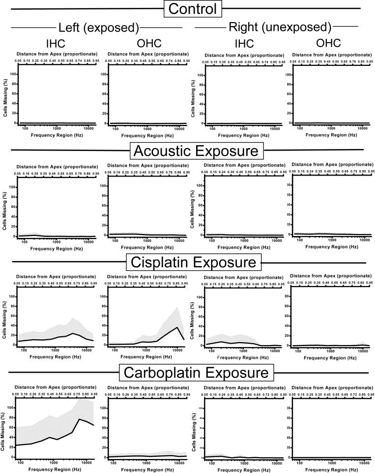
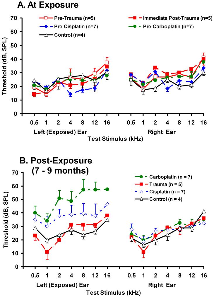
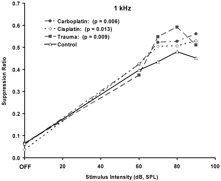
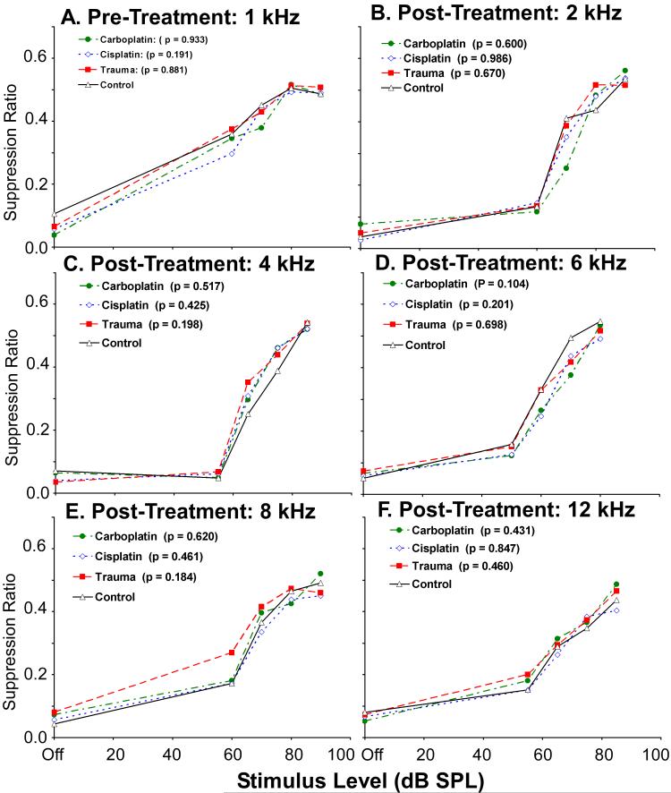
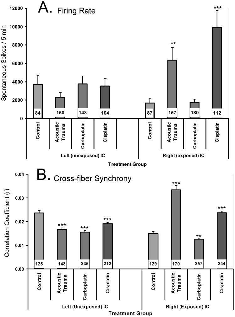
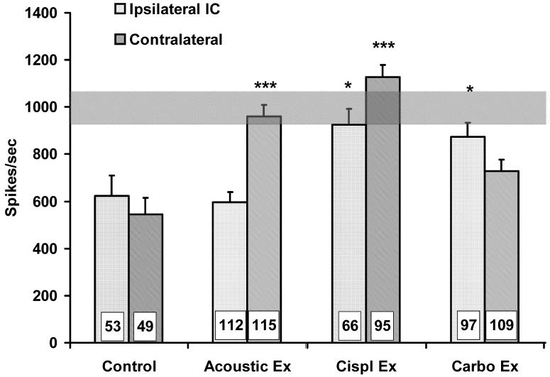
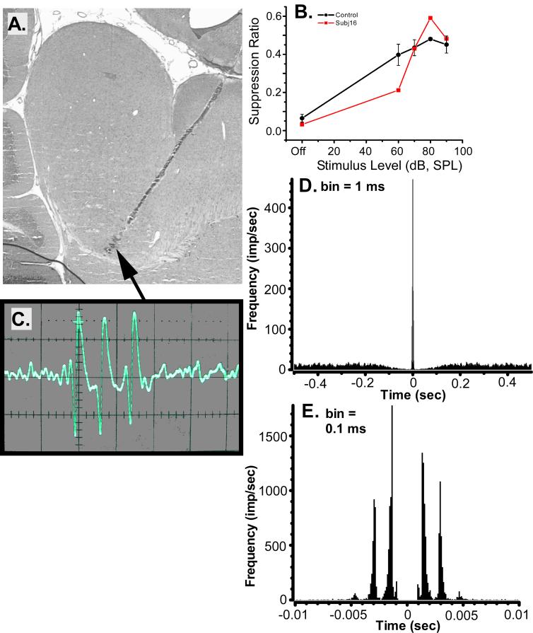
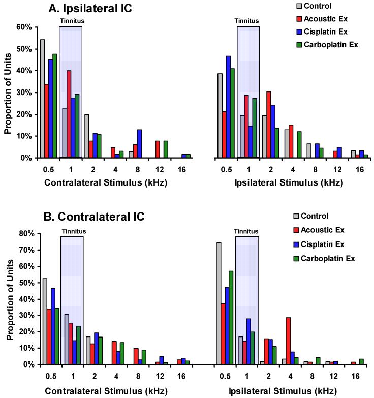
A longstanding hypothesis is that tinnitus, the perception of sound without an external acoustic source, is triggered by a distinctive pattern of cochlear hair cell (HC) damage and that this subsequently leads to altered neural activity in the central auditory pathway. This hypothesis was tested by assessing behavioral evidence of tinnitus and spontaneous neural activity in the inferior colliculus (IC) after unilateral cochlear trauma. Chinchillas were assigned to four cochlear treatment groups. Each treatment produced a distinctive pattern of HC damage, as follows: acoustic exposure (AEx): sparse low-frequency inner hair cell (IHC) and outer hair cell (OHC) loss; round window cisplatin (CisEx): pronounced OHC loss mixed with some IHC loss; round window carboplatin (CarbEx): pronounced IHC loss without OHC loss; control: no loss. Compared with controls, all experimental groups displayed significant and similar psychophysical evidence of tinnitus with features resembling a 1-kHz tone. Contralateral IC spontaneous activity was elevated in the AEx and CisEx groups, which showed increased spiking and increased cross-fiber synchrony. A multidimensional analysis identified a subpopulation of neurons more prevalent in animals with tinnitus. These units were characterized by high bursting, low ISI variance, and within-burst peak spiking of approximately 1,000/sec. It was concluded that cochlear trauma in general, rather than its specific features, leads to multiple changes in central activity that underpin tinnitus. Particularly affected was a subpopulation ensemble of IC neurons with the described unique triad of features.


Figures

Fig. 1
Fig. 1
A lesion-marked multi-probe electrode track in chinchilla IC, with superimposed image of a probe. Individual recording sites appear as punctuate lesions (numbered 1 - 16), separated by 100 μm. The separation between core and shell subdivisions is approximated by the curved broken line; the dorso-ventral partitioning of the IC is indicated by the horizontal broken lines. Sagittal view, fast H & E stain.
Fig.2
Fig.2
Average cytocochleograms of each treatment group, for both exposed and unexposed cochleas. The shaded area indicates + 1 standard deviation. Unexposed cochleas of all groups (two right-hand columns) showed little hair cell loss, with the exception of the cisplatin group in which some IHC loss was evident (on average less than 10 %). Control animals displayed normal cochleas, while acoustically exposed animals had less than 5 percent IHC and OHC loss in exposed cochleas. Cisplatin exposed animals showed both IHC and OHC loss in exposed cochleas. Carboplatin exposed animals showed a reasonably selective large loss of IHC in their exposed cochleas. IHC: inner hair cell; OHC: outer hair cell.
Fig.3
Fig.3
Acoustic brainstem evoked-response hearing thresholds obtained at the time of cochlear exposure (A) and at the time of electrophysiological data collection, 7 - 9 months after unilateral (left) exposure (B). Error bars show the standard error of the mean.
Fig.4
Fig.4
1 kHz tone discrimination indicative of tinnitus in the acoustic exposed (n = 5), cisplatin exposed (n = 7), and carboplatin exposed (n = 7) chinchillas, compared to unexposed controls (n = 4). The functions were determined 2 months after trauma or toxin exposure. The legend includes t statistic significance levels comparing experimental group functions (intensities > 0) to controls. Error bars indicate the standard error of the mean.
Fig.5
Fig.5
A. 1 kHz discrimination prior to cochlear insult. Error bars indicate the standard error of the mean. B. - F. Discrimination functions other than 1 kHz determined after cochlear insult, as depicted in Fig.4. Legends include t statistic significance levels comparing experimental group functions (intensities > 0) to controls.
Fig.6
Fig.6
Two features of spontaneous inferior colliculus single-neuron activity, obtained 8 - 9 months after unilateral exposure, of unexposed (n = 4), acoustic exposed (n = 5), cisplatin exposed (n = 7), and carboplatin exposed (n = 7) chinchillas. Recording epochs were 5 min. * p < 0.05, ** p < 0.01, *** p < 0.001. The number of single units contributing to each average is indicated by the number inset within each bar. Error bars indicate the standard error of the mean.
Fig.7
Fig.7
Scatter-plot depiction of spontaneous bursting as a function of single unit inter-spike-interval (ISI) variation for each of the treatment groups. Each data point represents a single unit. All units meeting the sorting criteria outlined in Electrophysiology: Data analysis are plotted. The solid line is an iterative least-mean square regression line. The vertical and horizontal broken lines index the inflection point of the regression line. The shaded area to the left of the inflection point encompasses the units used for the linear regression analysis summarized in Table 1. Left-column panels show results from the ipsilateral IC; right-column panels show results from the contralateral IC. The arrow (panel C) indicates data point for unit 16L2b, depicted in Fig. 9. There was no significant left and right difference in total spikes per 5 min, bursts per 5 min, mean peak frequency within burst, ISI (SD) or ISI mode for the control group.
Fig.8
Fig.8
Mean intra-burst peak spike frequency for the subpopulation of IC neurons falling within the shaded areas in Fig.7 is displayed for each group. The intra-burst peak frequency of the units in the exposed groups approximated the frequency at which the exposed animals showed psychophysical evidence of tinnitus (horizontal bar). All exposed groups were significantly different from controls, either contralaterally or bilaterally (* p < 0.05, *** p < 0.001). Error bars indicate the standard error of the mean (SEM). The horizontal gray bar represents ± 1 SEM for the unexposed control group.
Fig.9
Fig.9
An exemplary bursting unit, 16L2b, selected from an acoustic-exposed chinchilla with evidence of tinnitus. A. Sagittal IC section intersecting the track of the multiprobe site from which the unit was recorded. The arrow indicates the recording site of the unit. Distances between the lesion-marked recording sites were 100 μm. B. The psychophysical performance of subject 16 (square data points), from which unit 16L2b was obtained, compared to the performance of non-tinnitus control animals (circular data points, error bars represent the standard error of the mean). C. An oscilloscope trace of unit 16L2b showing its spontaneous burst pattern (minor tick marks: 0.5 msec). D. A low-resolution autocorrelogram of unit 16L2b, 1 sec window. E. A high-resolution autocorrelogram of unit 16L2b, 20 msec window.
Fig. 10
Fig. 10
Best frequency analysis of all confirmed recording sites. Multi-unit records were analyzed from the ipsilateral (A) and contralateral (B) IC for each treatment group. Best frequency was defined by the rate level function with the greatest stimulus-driven rate increase at asymptote. Both contralateral and ipsilateral pure tone stimuli were tested. There were no statistically significant differences between the control and experimental groups. Each panel summarizes the proportion of best frequencies for ipsilateral as well as contralateral stimulus driving.

References

    1. Alkhatib A, Biebel UW, Smolders JW. Reduction of inhibition in the inferior colliculus after inner hair cell loss. Neuroreport. 2006;17(14):1493–1497. - PubMed
    1. Atherley GRC, Hempstock TI, Noble WG. Study of tinnitus induced temporarily by noise. J Acoustical Soc Amer. 1968;44(6):1503–1506. - PubMed
    1. Bauer CA, Brozoski TJ. Assessing tinnitus and prospective tinnitus therapeutics using a psychophysical animal model. J of the Assoc for Res in Otolaryngol. 2001;2(1):54–64. - PMC - PubMed
    1. Bauer CA, Brozoski TJ. Cochlear structure and function after round window application of ototoxins. Hear Res. 2005;201(12):121–131. - PubMed
    1. Bohne BA. Location of small cochlear lesions by phase contrast microscopy prior to thin sectioning. Laryngoscope. 1972;82(1):1–16. - PubMed

Publication types