Review
. 2008 May;15(5):399-413.
doi: 10.1111/j.1553-2712.2008.00109.x.

Resuscitating the microcirculation in sepsis: the central role of nitric oxide, emerging concepts for novel therapies, and challenges for clinical trials

Collaborators, Affiliations
Review

Resuscitating the microcirculation in sepsis: the central role of nitric oxide, emerging concepts for novel therapies, and challenges for clinical trials

Stephen Trzeciak et al. Acad Emerg Med. 2008 May.

Abstract

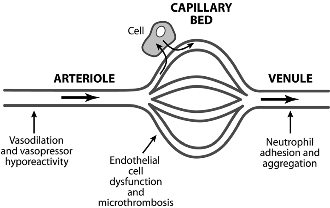
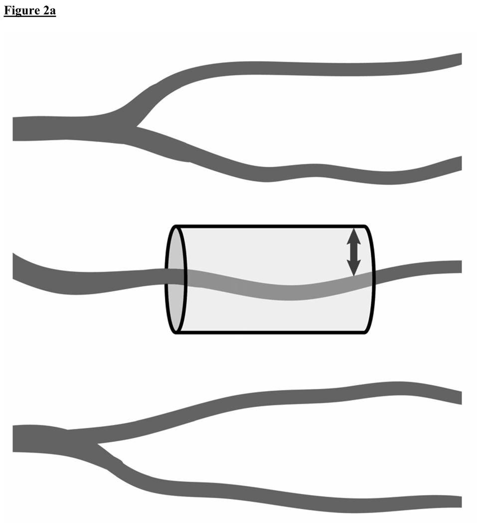
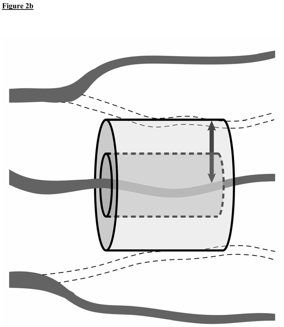
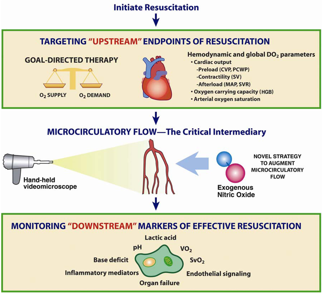
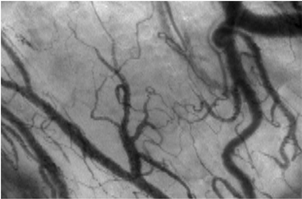
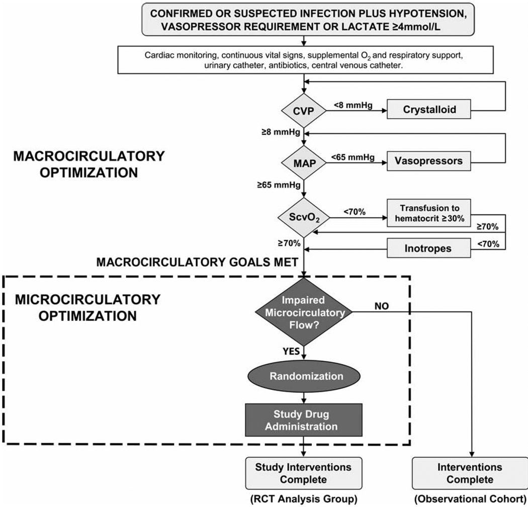
Microcirculatory dysfunction is a critical element of the pathogenesis of severe sepsis and septic shock. In this Bench-to-Bedside review, we present: 1) the central role of the microcirculation in the pathophysiology of sepsis; 2) new translational research techniques of in vivo video microscopy for assessment of microcirculatory flow in human subjects; 3) clinical investigations that reported associations between microcirculatory dysfunction and outcome in septic patients; 4) the potential role of novel agents to "rescue" the microcirculation in sepsis; 5) current challenges facing this emerging field of clinical investigation; and 6) a framework for the design of future clinical trials aimed to determine the impact of novel agents on microcirculatory flow and organ failure in patients with sepsis. We specifically focus this review on the central role and vital importance of the nitric oxide (NO) molecule in maintaining microcirculatory homeostasis and patency, especially when the microcirculation sustains an insult (as with sepsis). We also present the scientific rationale for clinical trials of exogenous NO administration to treat microcirculatory dysfunction and augment microcirculatory blood flow in early sepsis therapy.


Figures

Figure 1
Figure 1
Sepsis is a disorder of the microcirculation. Much of the pathophysiology of sepsis can be explained within the microcirculatory unit – the terminal arteriole, capillary bed, and the post-capillary venule. The arteriole is where the characteristic vasodilation and vasopressor hyporesponsiveness of sepsis occurs. The capillary bed is where the effects of endothelial cell activation/dysfunction are most pronounced and microvascular thromboses are formed. The post-capillary venule is where leukocyte trafficking is most disordered – leukocytes adhere to the vessel wall, aggregate, and further impair flow through the microcirculation.
Figure 2A and 2B
Figure 2A and 2B
A conceptual model of oxygen diffusion from capillaries. These figures illustrate how sepsis-induced microcirculatory dysfunction can play a key role in the impairment of tissue oxygen transport and contribute to tissue hypoxia. (2A)Healthy state: A cylinder represents the area of tissue that is supplied with oxygen by an individual capillary. The diffusion distance for oxygen in the tissues is shown (small arrow). (2B)Sepsis: Intrinsic microcirculatory dysfunction results in non-perfused capillaries (dotted line vessels). This decreases the density of perfused vessels, increasing the diffusion distance for oxygen in the tissues (large arrow).
Figure 2A and 2B
Figure 2A and 2B
A conceptual model of oxygen diffusion from capillaries. These figures illustrate how sepsis-induced microcirculatory dysfunction can play a key role in the impairment of tissue oxygen transport and contribute to tissue hypoxia. (2A)Healthy state: A cylinder represents the area of tissue that is supplied with oxygen by an individual capillary. The diffusion distance for oxygen in the tissues is shown (small arrow). (2B)Sepsis: Intrinsic microcirculatory dysfunction results in non-perfused capillaries (dotted line vessels). This decreases the density of perfused vessels, increasing the diffusion distance for oxygen in the tissues (large arrow).
Figure 3
Figure 3
Conceptual framework of the importance of the microcirculation in septic shock and resuscitation. Conventional resuscitation targets optimization of “upstream” (i.e. macrocirculatory) hemodynamic parameters (e.g. mean arterial pressure, cardiac output), with monitoring of “downstream” markers of tissue perfusion (e.g. acidosis, organ function) to determine the effectiveness of resuscitation efforts. The microcirculation represents a critical intermediary. Although the macrocirculation circulates blood throughout the body, an intact and functional microcirculation is necessary for effective blood flow to tissues. Therefore, intrinsic microcirculatory failure may contribute to sepsis-associated tissue hypoperfusion. Sublingual microcirculatory blood flow can now be visualized directly in sepsis clinical research using a hand-held videomicroscope (shown on left). In this paper, we present a scientific rationale for a clinical trial of a novel agent (e.g. exogenous nitric oxide administration, shown on right) to reduce microcirculatory dysfunction and augment microcirculatory blood flow in sepsis resuscitation. [CVP = central venous pressure; PCWP = pulmonary capillary wedge pressure; SV = stroke volume; MAP = mean arterial pressure; SVR = systemic vascular resistance; HGB = hemoglobin; VO2 = oxygen consumption; SvO2 = mixed venous oxygen saturation] Adapted from: Trzeciak S, Dellinger RP, Parrillo JE, Septic Shock, In: Parrillo JE and Dellinger RP (3rd Edition) Critical Care Medicine: Principles of Diagnosis and Management in the Adult. (2008) Philadelphia, PA: Mosby Elsevier.
Figure 4
Figure 4
A still image of the human sublingual microcirculation as visualized with Orthogonal Polarization Spectral (OPS) videomicroscopy. The videomicroscope uses a 5X objective (167X magnification) giving a 940 × 1259 µm field of view. Real-time video of healthy and dysfunctional microcirculation is available for viewing or download at: http://www.cooperhealth.org/content/gme_fellowship_shock.htm
Figure 5
Figure 5
A template for designing a randomized clinical trial of a novel agent to augment microcirculatory flow and improve outcome in sepsis resuscitation. Microcirculatory flow would be assessed with in vivo videomicroscopy at the bedside. Although clinically speaking, an agent that improves microcirculatory flow might optimally be initiated immediately at the time of severe sepsis identification, a requisite for this type of clinical trial would be early achievement of homogeneity in macrocirculatory hemodynamic optimization (e.g. early goal-directed therapy as per Rivers et al 4 or a similar resuscitation algorithm) in both the control and treatment subjects, in order to permit precise determination of the treatment effect of microcirculatory optimization on outcome. Because enrolling patients who do not manifest the microcirculatory dysfunction phenotype could cause the clinical trial to be underpowered to show a treatment effect, we advocate a “personalized” trial design employing a real-time assessment of microcirculatory flow prior to the decision to randomize (as shown). [CVP = central venous pressure; MAP = mean arterial pressure; ScvO2 = central venous oxygen saturation; RCT = randomized controlled trial]

References

    1. Angus DC, Linde-Zwirble WT, Lidicker J, Clermont G, Carcillo J, Pinsky MR. Epidemiology of severe sepsis in the United States: analysis of incidence, outcome, and associated costs of care. Crit Care Med. 2001 Jul;29(7):1303–1310. - PubMed
    1. Hotchkiss RS, Karl IE. The pathophysiology and treatment of sepsis. N Engl J Med. 2003 Jan 9;348(2):138–150. - PubMed
    1. Kumar A, Roberts D, Wood KE, et al. Duration of hypotension before initiation of effective antimicrobial therapy is the critical determinant of survival in human septic shock. Crit Care Med. 2006 Jun;34(6):1589–1596. - PubMed
    1. Rivers E, Nguyen B, Havstad S, et al. Early goal-directed therapy in the treatment of severe sepsis and septic shock. N Engl J Med. 2001 Nov 8;345(19):1368–1377. - PubMed
    1. Lin SM, Huang CD, Lin HC, Liu CY, Wang CH, Kuo HP. A modified goal-directed protocol improves clinical outcomes in intensive care unit patients with septic shock: a randomized controlled trial. Shock. 2006 Dec;26(6):551–557. - PubMed

Publication types