. 2008 Apr 24;58(2):273-83.
doi: 10.1016/j.neuron.2008.01.025.

Know your place: neural processing of social hierarchy in humans

Affiliations

Know your place: neural processing of social hierarchy in humans

Caroline F Zink et al. Neuron. .

Abstract

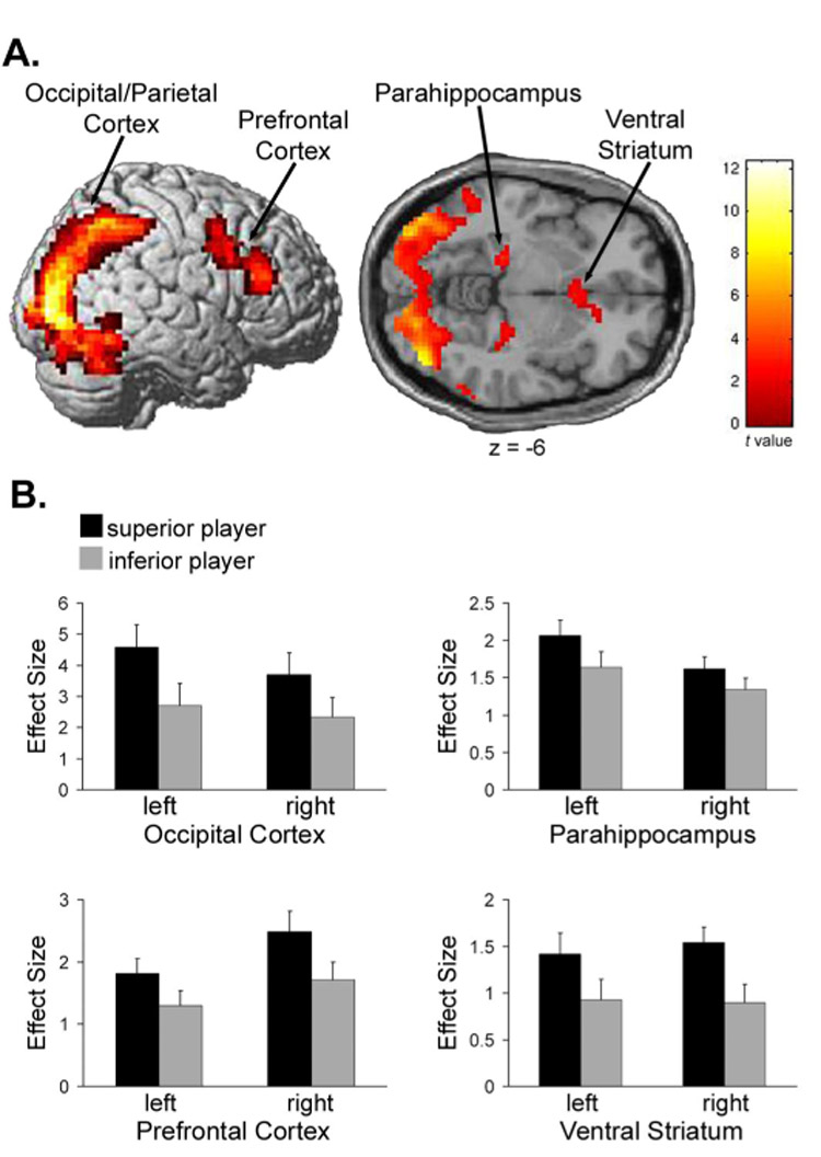
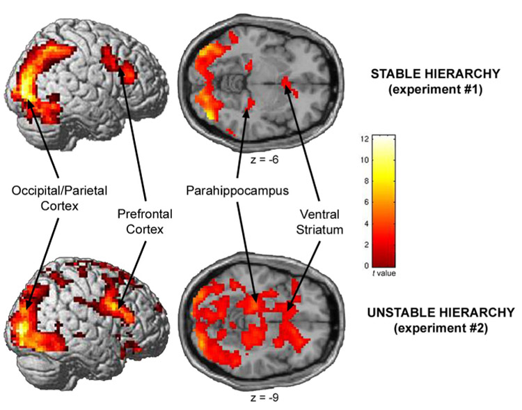
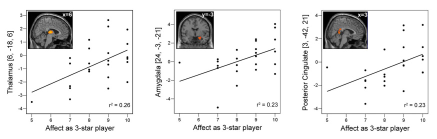
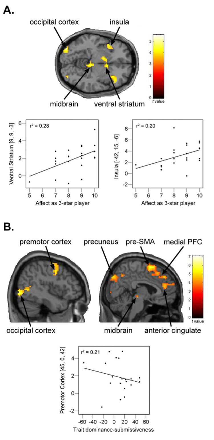
Social hierarchies guide behavior in many species, including humans, where status also has an enormous impact on motivation and health. However, little is known about the underlying neural representation of social hierarchies in humans. In the present study, we identify dissociable neural responses to perceived social rank using functional magnetic resonance imaging (fMRI) in an interactive, simulated social context. In both stable and unstable social hierarchies, viewing a superior individual differentially engaged perceptual-attentional, saliency, and cognitive systems, notably dorsolateral prefrontal cortex. In the unstable hierarchy setting, additional regions related to emotional processing (amygdala), social cognition (medial prefrontal cortex), and behavioral readiness were recruited. Furthermore, social hierarchical consequences of performance were neurally dissociable and of comparable salience to monetary reward, providing a neural basis for the high motivational value of status. Our results identify neural mechanisms that may mediate the enormous influence of social status on human behavior and health.


Figures

Figure 1
Figure 1
Schematic diagram of the experimental design. Each round in the tasks consisted of three phases: viewing the other player, playing the game, and viewing the outcomes. (A) In Experiment #1 (stable hierarchy), during the game phase participants pressed a button as soon as the blue circle changed to green. The initial hierarchical rankings did not change throughout the session. (B) In Experiment #2 (unstable hierarchy), during the game phase participants pressed a button to indicate which box contained more dots. The hierarchical rankings were updated throughout the session based on performance.
Figure 2
Figure 2
Significant activations for the contrast, “superior player > inferior player” in Experiment #1 (stable hierarchy). Displayed are (A) significant (P < 0.005, FDR-corrected) activations in occipital/parietal cortex [−24, −96, 9; 42, −81, −6], dorsal lateral prefrontal cortex [−36, 3, 42; 42, 30, 21], parahippocampal cortex [−21, −27, −9; 27, −24, −12], and ventral striatum [−3, 15, −6; 6, 18, −3], and (B) plots of the effect sizes (parameter estimates) when viewing the superior and inferior other player, extracted from the peak voxels in each activated region.
Figure 3
Figure 3
Significant activations for the contrast, “superior player > inferior player”, consistently observed in both Experiment #1 (top) and Experiment #2 (bottom). Significant (P < 0.005, FDR-corrected) activations were observed in occipital/parietal cortex (Experiment #1: [−24, −96, 9; 42, −81, −6]; Experiment #2: [−27, −93, 6; 36, −93, 3]), 35 dorsal lateral prefrontal cortex (Experiment #1: [−36, 3, 42; 42, 30, 21]; Experiment #2: [45, 9, 27]), parahippocampal cortex (Experiment #1: [−21, −27, −9; 27, −24, −12]; Experiment #2: [−24, −27, −12; 27, −21, −15]), and ventral striatum (Experiment #1: [−3, 15, −6; 6, 18, −3]; Experiment #2: [9, 9, −3]).
Figure 4
Figure 4
Correlations between brain activity and the level of positive affect experienced by the participant when in the top hierarchical position in areas uniquely activated in Experiment #2 for the contrast, “superior player > inferior player”. Displayed are significant (P < 0.05) correlations between the level of positive affect experienced by the participant as the three star player and parameter estimates at peak activations when viewing the superior player in the thalamus (P = 0.011; r = 0.51), amygdala (P = 0.017; r = 0.481) and posterior cingulate (P = 0.018; r = 0.478).
Figure 5
Figure 5
Significant activations to outcomes associated with hierarchical value in Experiment #2, displayed at P < 0.001, uncorrected. (A) Activations for the contrast, “subject lost/inferior won > subject lost/inferior lost”, observed in occipital cortex [−36, − 90, −6; 33, −87, 9], insula [−42, 15, −6; 36, 24, 6], midbrain [−3, −30, −12], and ventral straitum [−6, 6, −3; 9, 9, −3]. Also displayed are significant correlations between the level of positive affect experienced by the participant as the three star player and parameter estimates at peak activations when subject lost/inferior won in the ventral striatum (P = 0.008; r = 0.528) and insula (P = 0.030; r = 0.444). (B) Activations for the contrast, “subject won/superior lost > subject won/superior won”, observed in occipital cortex [− 48, −72, 0; 45, −84, −6], premotor cortex [−39, −6, 57; 45, 0, 42], precuneus [−9, −81, 45; −24, −78, 33; 21, −87, 42], midbrain [−3, −21, −21; 9, −24, −6], pre-supplementary motor area [3, 9, 63], medial prefrontal cortex [3, 42, 39], and anterior cingulate [9, 42, 18]. Not shown are activations in orbitofrontal cortex [33, 21, −18] and caudate [−6, 6, 9]. Also displayed are significant negative correlations between trait dominance/submissiveness scores and parameter estimates at peak activations when subject won/superior lost in the premotor cortex (P = 0.04; r = −0.453).

Comment in

Similar articles

Cited by

References

    1. Abler B, Walter H, Erk S. Neural correlates of frustration. Neuroreport. 2005;16:669–672. - PubMed
    1. Adolphs R. Is the human amygdala specialized for processing social information? Ann. N.Y. Acad. Sci. 2003;985:326–340. - PubMed
    1. Aminoff E, Gronau N, Bar M. The parahippocampal cortex mediates spatial and nonspatial associations. Cereb. Cortex. 2007;17:1493–1503. - PubMed
    1. Amodio DM, Frith CD. Meeting of minds: the medial frontal cortex and social cognition. Nat. Rev. Neurosci. 2006;7:268–277. - PubMed
    1. Ashburner J, Friston KJ. Nonlinear spatial normalization using basis functions. Hum. Brain Mapp. 1999;7:254–266. - PMC - PubMed

Publication types

LinkOut - more resources