. 2008 Jun;38(2):125-37.
doi: 10.1016/j.mcn.2008.01.014. Epub 2008 Feb 13.

Loss of c/EBP-beta activity promotes the adaptive to apoptotic switch in hypoxic cortical neurons

Affiliations

Loss of c/EBP-beta activity promotes the adaptive to apoptotic switch in hypoxic cortical neurons

Marc W Halterman et al. Mol Cell Neurosci. 2008 Jun.

Abstract

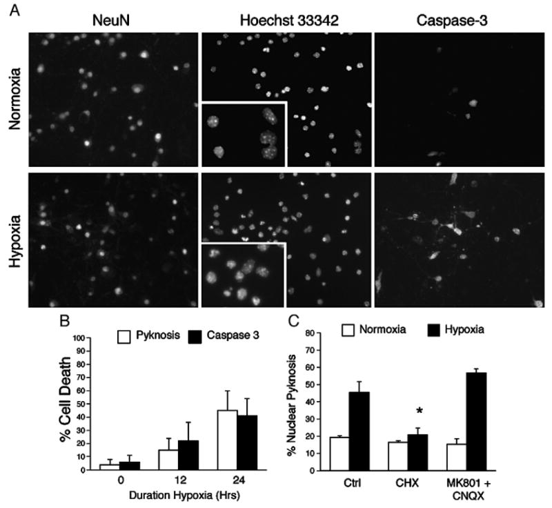
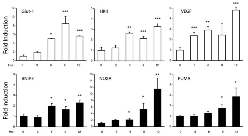
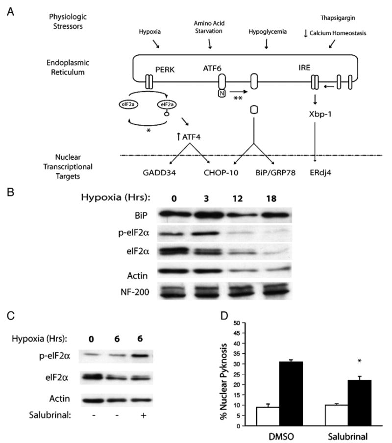
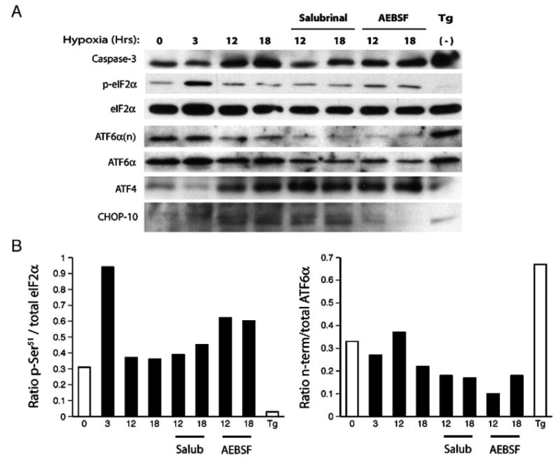
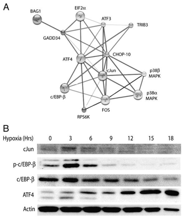
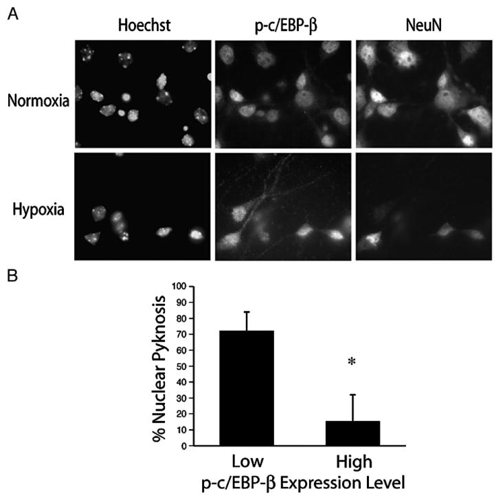
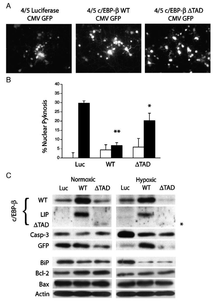
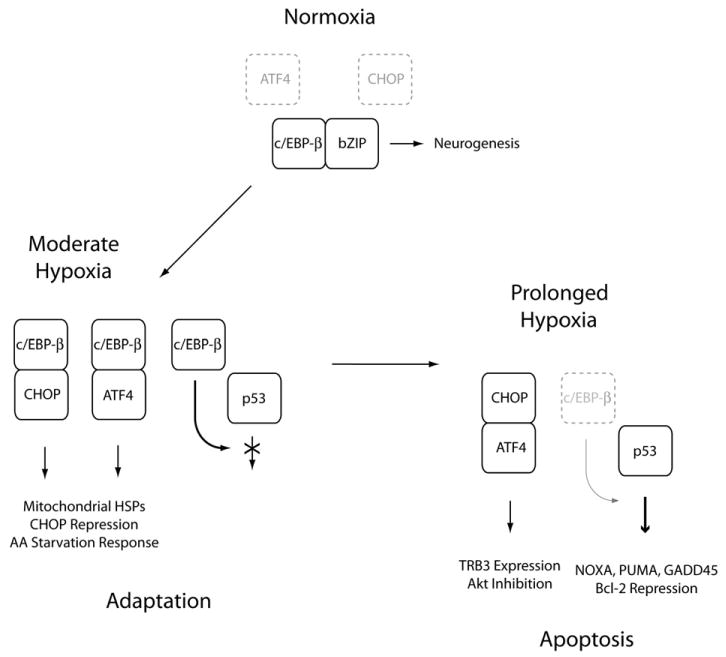
Understanding the mechanisms governing the switch between hypoxia-induced adaptive and pathological transcription may reveal novel therapeutic targets for stroke. Using an in vitro hypoxia model that temporally separates these divergent responses, we found apoptotic signaling was preceded by a decline in c/EBP-beta activity and was associated with markers of ER-stress including transient eIF2alpha phosphorylation, and the delayed induction of the bZIP proteins ATF4 and CHOP-10. Pretreatment with the eIF2alpha phosphatase inhibitor salubrinal blocked the activation of caspase-3, indicating that ER-related stress responses are integral to this transition. Delivery of either full-length, or a transcriptionally inactive form of c/EBP-beta protected cultures from hypoxic challenge, in part by inducing levels of the anti-apoptotic protein Bcl-2. These data indicate that the pathologic response in cortical neurons induced by hypoxia involves both the loss of c/EBP-beta-mediated survival signals and activation of pro-death pathways originating from the endoplasmic reticulum.


Conflict of interest statement

The authors report no commercial affiliations or conflicts of interest pertaining to this manuscript.

Figures

Fig. 1
Fig. 1
Chronic hypoxia activates apoptosis in dissociated neuronal cultures. (A) Continuous hypoxia induces nuclear pyknosis and caspase-3 cleavage in DIV7 cortical neurons. Neuronal cultures were exposed to hypoxia (0.5% O2) for 18 h and analyzed by fluorescence microscopy for expression of the neuronal marker NeuN, nuclear pyknosis using the DNA dye Hoechst 33342, and the cleavage of caspase-3. (B) Nuclear pyknosis approximates caspase-3 cleavage in vitro. (C) Hypoxia-induced neuron death requires de novo translation. Cultures were pretreated with cycloheximide (CHX 1 μg/ml, 3.5 μM) or a combination of the glutamate receptor antagonists MK-801 (100 μM) and CNQX (10 μM), and exposed hypoxia (0.5% O2, 18 h; filled bars) and analyzed for nuclear pyknosis versus control (21% O2; open bars) sister cultures. Data represent the average±S.D. of results from six non-overlapping fields across replicates (n=4; *=P<0.05).
Fig. 2
Fig. 2
Transcriptional profiling by quantitative RT-PCR defines adaptive and pathologic phases of gene expression. Neuronal cultures were made hypoxic for 3, 6, 9 and 12 h, total RNA was harvested and analyzed by quantitative PCR for the adaptive targets vascular endothelial growth factor (VEGF), hexokinase II (HKII) and the glucose transporter (Glut-1) (open bars), or the pro-apoptotic factors BNIP3, NOXA and PUMA (filled bars). Data are expressed as fold-induction relative to normoxic samples and expressed as the average±S.D. of results from sister wells (n=3; *=P<0.05, **=P<0.01, ***=P<0.001).
Fig. 3
Fig. 3
The unfolded protein response regulates neuron survival after chronic hypoxia. (A) Schematic representation of the physiologic stressors associated with ischemic stress, the proximate shared (eIF2α) and specific (ATF6, IRE/Xbp-1) ER sensors responsive to these cues and their downstream nuclear transcriptional targets. The sites of action of the pharmacological inhibitors salubrinal (*) and the serine protease inhibitor AEBSF (**) are indicated. (B) Western analysis indicates that hypoxia stimulates the transient induction of BiP/GRP78 and phosphorylation of eIF2α. Pretreatment with the phosphatase inhibitor salubrinal (10 μM) enhances eIF2α phosphorylation in hypoxic neurons (C), and (D) limits hypoxia-induced neuronal pyknosis in vitro (18 h, 0.5% O2; filled bars). Data for control (open bars) and hypoxic samples and expressed as the average±S.D. (n=3 wells; *=P<0.01).
Fig. 4
Fig. 4
Hypoxia-induced ER-stress responses regulate neuron survival and the delayed expression of the bZIP factors ATF4 and CHOP-10. (A) Protein lysates were harvested from normoxic (0 Hr) or hypoxic cultures (3–18 Hrs; 0.5% O2) and analyzed by western blotting for caspase-3 cleavage, eIF2α phosphorylation, proteolytic cleavage of ATF6, release of the transcriptionally active ATF6 amino-terminus (ATF6α(n)), and induction of the bZIP proteins ATF4 and CHOP-10. Sister cultures were also treated with salubrinal (10 μM), the serine protease inhibitor AEBSF (100 μM) or the Ca2+-ATPase inhibitor thapsigargin (Tg; 1 μg/ml) and exposed to hypoxia (excluding Tg treated samples) as indicated. (B) Western blots were quantified using densitometry and the data are expressed histogram form as the ratios of p-eIF2α/total eIF2α and ATF6α(n)/total ATF6α.
Fig. 5
Fig. 5
Dynamic regulation of the bZIP family of transcription factors in hypoxic neurons. (A) The STRING protein–protein interaction algorithm (http://string.embl.de/) implicates the bZIP factors c/EBP-β and c-Jun in the regulated activity of ATF4 and CHOP-10. (B) Western analysis of hypoxic neuronal cultures for c-Jun, total and phosphorylated c/EBP-β (Thr188).
Fig. 6
Fig. 6
Persistent expression of phosphorylated c/EBP-β Thr188 is a marker for neuron survival following hypoxic stress. (A) Dissociated cortical cultures were exposed to hypoxia (18 h, 0.5% O2) and analyzed by ICC for c/EBP-β Thr188 and the neuronal marker NeuN. (B) Nuclear morphology was determined for neurons expressing either high or low levels of p-c/EBP-β as indicated. Data represent the average±S.D. of results from five non-overlapping fields (*=P<0.05).
Fig. 7
Fig. 7
Viral-mediated expression of c/EBP-β supports neuron survival after hypoxic challenge. (A) The bi-cistronic herpes amplicon vector HSVprPucCMVeGFP identifies neurons expressing luciferase (LUC), full-length c/EBP-β (WT), or a truncated form of c/EBP-β (ΔTAD). (B) c/EBP-β expression promotes neuron survival after hypoxic challenge. DIV7 cultures were transduced with the expression constructs as indicated and analyzed for levels of nuclear pyknosis after 18 h of normoxic (open bars) or hypoxic (closed bars) conditions. The data represent the average (±S.D.) pyknosis amongst transduced neurons from five non-overlapping fields (n=3; *=P<0.05, **=P<0.01). (C) Expression of full-length or truncated c/EBP-β (ΔTAD) limit hypoxia-induced caspase-3 cleavage and enhance Bcl-2 levels. Cortical neurons were transduced with wild-type, dominant-negative (ΔTAD) and control virus 12 h prior to hypoxic challenge and subjected to 18 h of hypoxia (0.5% O2). Lysates were harvested and analyzed by western blotting for the expression of the full-length, internally translated inhibitory fragment (LIP), the ΔTAD fragment (asterisk), activated caspase-3, GFP, BiP/GRP78, Bcl-2, and Bax.
Fig. 8
Fig. 8
Hypoxia-mediated loss of neuronal c/EBP-β activity promotes the adaptive to apoptotic transition. This schematic illustrates our conceptual model of how attenuated c/EBP-β activity promotes the adaptive to apoptotic transition in hypoxic neurons. Under normoxic conditions, c/EBP-β associates with other neuronal bZIP heterodimeric partners to regulate neurogenesis. Under conditions of sub-lethal hypoxic stress, ER-stress induces the expression of ATF4 and CHOP-10, which associate with c/EBP-β to drive the expression of mitochondrial heat shock proteins and genes involved in the amino acid (AA) starvation response. c/EBP-β also functions as an inhibitory factor repressing the expression of CHOP-10 as well as directly trans-repressing the expression of pro-apoptotic p53 targets. Under conditions of severe hypoxic stress, c/EBP-β levels decline allowing for the formation of pathological bZIP heterodimers (i.e., ATF4:CHOP-10) and the activation of p53-dependent latent pro-apoptotic transcriptional activity.

References

    1. Abe K, Aoki M, Kawagoe J, Yoshida T, Hattori A, Kogure K, Itoyama Y. Ischemic delayed neuronal death. A mitochondrial hypothesis. Stroke. 1995;26:1478–1489. - PubMed
    1. Aldridge JE, Horibe T, Hoogenraad NJ. Discovery of genes activated by the mitochondrial unfolded protein response (mtUPR) and cognate promoter elements. PLoS ONE. 2007;2:e874. - PMC - PubMed
    1. Ankarcrona M, Dypbukt JM, Bonfoco E, Zhivotovsky B, Orrenius S, Lipton SA, Nicotera P. Glutamate-induced neuronal death: a succession of necrosis or apoptosis depending on mitochondrial function. Neuron. 1995;15:961–973. - PubMed
    1. Banasiak KJ, Haddad GG. Hypoxia-induced apoptosis: effect of hypoxic severity and role of p53 in neuronal cell death. Brain Res. 1998;797:295–304. - PubMed
    1. Blais JD, Filipenko V, Bi M, Harding HP, Ron D, Koumenis C, Wouters BG, Bell JC. Activating transcription factor 4 is translationally regulated by hypoxic stress. Mol Cell Biol. 2004;24:7469–7482. - PMC - PubMed

Publication types

MeSH terms

Substances