. 2008 Apr 25;30(2):145-55.
doi: 10.1016/j.molcel.2008.02.023.

Architecture of a serine recombinase-DNA regulatory complex

Affiliations

Architecture of a serine recombinase-DNA regulatory complex

Kent W Mouw et al. Mol Cell. .

Abstract

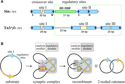
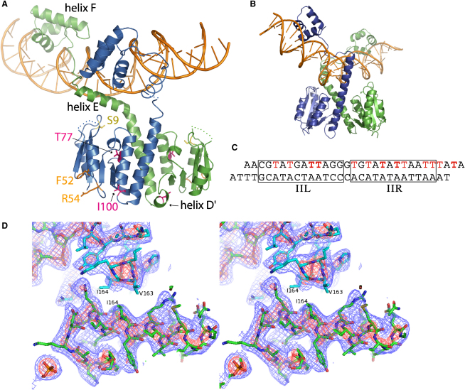
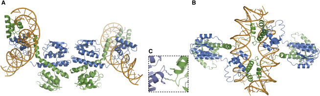
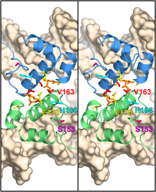
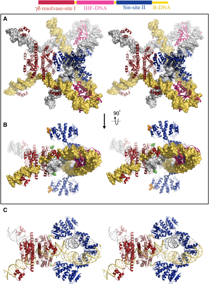
An essential feature of many site-specific recombination systems is their ability to regulate the direction and topology of recombination. Resolvases from the serine recombinase family assemble an interwound synaptic complex that harnesses negative supercoiling to drive the forward reaction and promote recombination between properly oriented sites. To better understand the interplay of catalytic and regulatory functions within these synaptic complexes, we have solved the structure of the regulatory site synapse in the Sin resolvase system. It reveals an unexpected synaptic interface between helix-turn-helix DNA-binding domains that is also highlighted in a screen for synapsis mutants. The tetramer defined by this interface provides the foundation for a robust model of the synaptic complex, assembled entirely from available crystal structures, that gives insight into how the catalytic activity of Sin and other serine recombinases may be regulated.


Figures

Figure 1
Figure 1
res Site Architecture and Recombination Topology (A) res site architecture. In the Sin system, each res site binds two dimers of Sin and an architectural protein such as HU or IHF (at the synthetic resF site). The res sites from the Tn3/γδ systems are longer than the Sin res site (114 versus 86 bp) and bind three separate recombinase dimers and no architectural proteins. In both systems, DNA cleavage occurs within site I to generate 2 nt 3′ overhangs, while the regulatory sites (site II and the HU/IHF binding site in the Sin system and sites II and III in the Tn3/γδ systems) serve to stimulate recombination and direct the outcome of the reaction. (B) Recombination topology. Sin resolves plasmid dimers to monomers by recombining two res sites that are present in direct repeat on a supercoiled plasmid. The synaptic complexes assembled by Sin and by Tn3/γδ resolvase trap three (−) interdomainal supercoils, and the predominant reaction product is a two-noded catenane.
Figure 2
Figure 2
Crystal Structure of the Site II-Bound Sin Dimer (A) Individual Sin monomers are shown in blue and green; the site II duplex is in orange. Ser-9, the active site nucleophile at site I, is shown in yellow. The positions of activating (T77I, I100T) and regulatory (F52, R54) mutations highlighted in the genetic screens (see Figure 5) are shown in magenta and orange, respectively. Residues 35–41 from both monomers, as well as residues 130 and 131 from the blue monomer, are not present in the final model but are represented here with dotted lines. (B) Structure of the γδ resolvase site I dimer complex (Yang and Steitz, 1995). (C) The sequence of the site II duplex in the crystal. The left and right half-sites of the site II direct repeat are boxed. T's shown in red were substituted with 5-Br-dU, and bold T's were substituted with 5-I-dU. (D) Stereo-view experimental electron density map showing interface between the CTDs of adjacent Sin dimers in the crystal. Residues V163 and I164 are at the center of the interface (see text). The maps contain no model phase information and are contoured at 2.5 σ (red) and 1.0 σ (blue).
Figure 3
Figure 3
Tetrameric Interfaces Observed in the Crystal Structure (A) An interface involving the N-terminal catalytic domains places the bound site II duplexes along the outside of the complex. The duplexes define a right-handed (+) node crossing; this structure is thus a poor candidate for the site II synaptic complex. (B) The interface between DNA-binding domains defines a tetramer in which the bound duplexes are near the center of the complex and cross to form a left-handed (−) node. The orientation and close proximity of the duplexes make this a good candidate for the site II synaptic interface. (C) A close-up view of the interdigitating interaction involving the side chains of residues F52 and R54 from two adjacent dimer complexes in the crystal structure (see also Figure S1).
Figure 4
Figure 4
Stereo View of the Interface between Sin DNA-Binding Domains in the Site II Synaptic Tetramer Residues from helix F comprise much of the interface. Side chains are shown for all residues that, when mutated, confer a defect in synapsis and recombination (see Figure 5). Red, V163 and I164; orange, Q160, K161, and R167; yellow, E170 and N186; and magenta, S153. Also shown is H166 (cyan), the position of the suppressor mutation H166R. The DNA-binding domains of Sin subunits bound at site IIL (green) and site IIR (blue) are shown (N-terminal domains not shown).
Figure 5
Figure 5
Effects of CTD Synapsis Mutations and R54E on the Regulation of Sin Recombination (A) Substrate plasmid used to select site II synapsis mutants in vivo, and site II synapsis assays for WT Sin and I164T. Site II synapsis by WT Sin blocks Cre-loxP recombination, preventing loss of the galK gene (red colonies on indicator plates); synapsis mutants (e.g., I164T) fail to block Cre (white colonies). (B) Quantitative recombination assays. The listed mutations were selected as causing a defect in site II synapsis in a WT background. The effect of each mutation on recombination is shown in a WT background (after ∼57 generations of growth) and in an I100T background (after ∼28 generations). Note that values of 100% recombination, seen in the I100T background, represent saturation of the assay. (C) H166R was selected as a second-site mutation that suppresses the inhibitory effect of S153T on res × res recombination in an I100T background. White colonies indicate that most or all of the test substrate has been resolved after ∼20 generations of growth; red colonies indicate inhibition of recombination. (D) The mutation R54E selectively inhibits res × res recombination, and the effect can be suppressed by a CTD mutation (e.g., I164T). Recombination was assayed in a T77I background; T77I is an activated mutant of Sin that can recombine site I × site I substrates (S.J.R., unpublished data). Inhibition by R54E requires site II in both recombining sites (data not shown), suggesting that site II synapsis is required.
Figure 6
Figure 6
A Model for the Sin Synaptic Complex in Stereo View (A) The model was constructed by rigid body docking of the Sin-site II synaptic tetramer (Figure 3B) with existing crystal structures of the IHF-DNA (Rice et al., 1996) and γδ resolvase-site I tetramer (Li et al., 2005) complexes. A 12 bp segment of canonical B form DNA has been added at the end of each site II in order to better visualize the path of the DNA as it exits the synaptic complex. No steric clashes between protein subunits are observed, and the DNA forms the three (−) nodes predicted by topological experiments. The position and orientation of the IHF site are as in resFD (Rowland et al., 2006). (B) A second view of the synaptic complex model rotated 90° about a horizontal axis. Residues implicated in intertetramer communication are highlighted: Sin F52 and R54 at site II are shown in orange, and the equivalent positions in γδ resolvase at site I (residues 54 and 56) are shown in green. (C) A direct interaction between Sin bound at sites I and II can be modeled by using the observed crystallographic interface involving residues F52 and R54 (Figure 3C). The NTDs of the site I-bound γδ tetramer have been replaced with Sin NTDs, and IHF-DNA complexes have been removed from the model for clarity. Sin residues F52 and R54 from sites I and II are shown in green and orange, respectively. Unlike the model in (A) and (B), adjustments to the protein are required to construct this model and result in small gaps in the protein (at the hinge region of the site II-bound Sin dimers, shown in pink) and the DNA (the spacer bound by IHF, not shown, does not completely bridge the site I–site II gap).

Comment in

References

    1. Biswas T., Aihara H., Radman-Livaja M., Filman D., Landy A., Ellenberger T. A structural basis for allosteric control of DNA recombination by λ integrase. Nature. 2005;435:1059–1066. - PMC - PubMed
    1. Bregu M., Sherratt D.J., Colloms S.D. Accessory factors determine the order of strand exchange in Xer recombination at psi. EMBO J. 2002;21:3888–3897. - PMC - PubMed
    1. Brunger A.T., Adams P.D., Clore G.D., DeLano W.L., Gros P., Grosse-Kunstleve R.W., Jiang J.-S., Kuszewski J., Nilges M., Pannu N.S. Crystallography & NMR system: a new software suite for macromolecular structure determination. Acta Crystallogr. D Biol. Crystallogr. 1998;54:905–921. - PubMed
    1. Calos M.P. The ϕC31 integrase system for gene therapy. Curr. Gene Ther. 2006;6:633–645. - PubMed
    1. Canosa I., Lopez G., Rojo F., Boocock M.R., Alonso J.C. Synapsis and strand exchange in the resolution and DNA inversion reactions catalyzed by the β recombinase. Nucleic Acids Res. 2003;31:1038–1044. - PMC - PubMed

Publication types

Associated data