. 2008 Apr 25;30(2):248-58.
doi: 10.1016/j.molcel.2008.02.021.

Toward a comprehensive temperature-sensitive mutant repository of the essential genes of Saccharomyces cerevisiae

Affiliations

Toward a comprehensive temperature-sensitive mutant repository of the essential genes of Saccharomyces cerevisiae

Shay Ben-Aroya et al. Mol Cell. .

Abstract

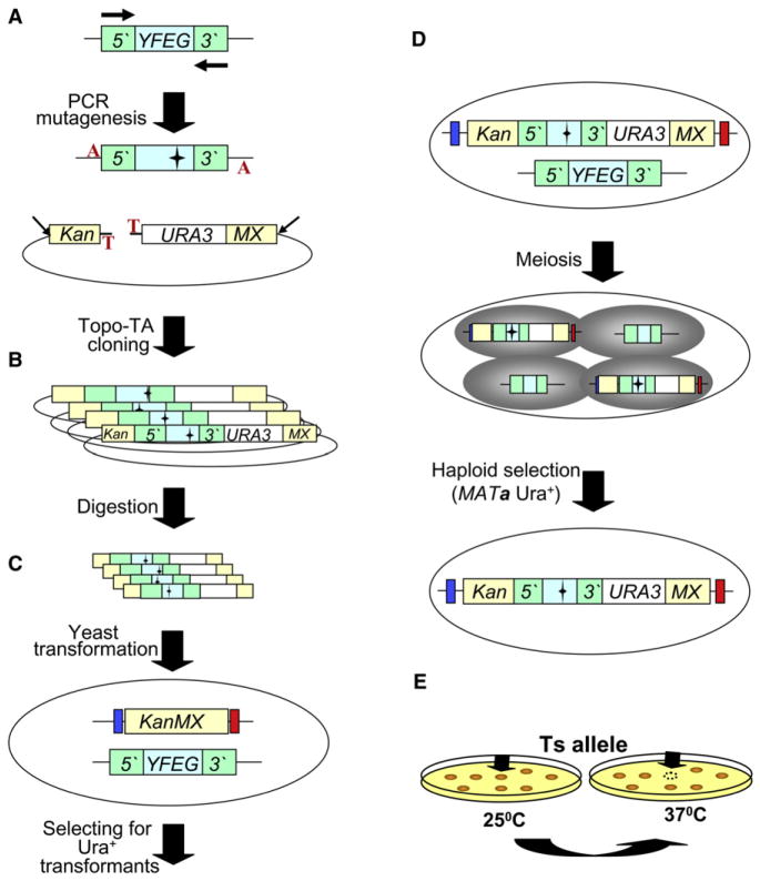
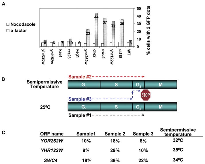
The Saccharomyces cerevisiae gene deletion project revealed that approximately 20% of yeast genes are required for viability. The analysis of essential genes traditionally relies on conditional mutants, typically temperature-sensitive (ts) alleles. We developed a systematic approach (termed "diploid shuffle") useful for generating a ts allele for each essential gene in S. cerevisiae and for improved genetic manipulation of mutant alleles and gene constructs in general. Importantly, each ts allele resides at its normal genomic locus, flanked by specific cognate UPTAG and DNTAG bar codes. A subset of 250 ts mutants, including ts alleles for all uncharacterized essential genes and prioritized for genes with human counterparts, is now ready for distribution. The importance of this collection is demonstrated by biochemical and genetic screens that reveal essential genes involved in RNA processing and maintenance of chromosomal stability.


Figures

Figure 1
Figure 1. Diploid Shuffle—A Method for Generating ts Alleles for the Essential Genes in Saccharomyces cereviviae
(A) Genomic DNA containing YFEG and its 5′ and 3′ regions is used as the template for PCR mutagenesis. Two black horizontal arrows represent the gene-specific primers used. The mutagenized PCR product is cloned into the vector SB221+ Topo-TA (mutations are represented by black stars). The Topo-TA cloning site is represented by a red T, and the A overhang protruding from the PCR product is represented by a red A. Left beige bar represents the 5′ half of the KanMX selectable marker (Kan), while the right beige bar represents the other half of the KanMX selectable marker (MX). The NotI restriction sites are indicated by two diagonal black arrows. (B) The product of the cloning step is a library of a mutagenized YFEG. The library is then transformed into E. coli and digested with NotI to release linear fragments (following DNA purification). (C) The linearized library is transformed into the corresponding heterozygous diploid strain. Blue and red bars that flank the KanMX knockout represent the two bar codes. (D) Heterozygous diploid transformants are sporulated (following meiosis), and MATa Ura+ haploids spores are selected on haploid-selective medium at 25°C. (E) Selection of ts candidates following the replica plating and incubating at 25°C and 37°C. Black arrows identify a potential ts allele.
Figure 2
Figure 2. Ribosomal RNA-Processing Screen
(A) Scheme of rRNA processing. Ribosomal RNA is encoded as part of ~9 kb tandem repeat in yeast and is processed from a 35S precursor in the multistep pathway outlined here. The cartoon indicates rDNA repeat structure and positions of mature 25S, 5.8S, and 18S rRNAs (black bars in DNA and precursor forms, gray bars represent mature forms) derived from the precursor, as well as the separately transcribed 5S rRNA. 3′ ends are indicated by arrowheads. Intermediates and pathways of rRNA processing have been identified and are shown in the bottom part of the panel. Dotted lines indicate effects of processing; an alternate pathway to the formation of mature 5.8S and 25S rRNAs from the topmost 27S precursor is indicated by square bracket. Note that there are multiple species with mobilities of about 27S and 7S; our experiments do not distinguish among these. The internal transcribed spacer (ITS) regions used as probes to detect precursor forms are indicated in the inset. (B) Isolation of ribosomal RNA-processing-defective mutants. RNA preparations were resolved on 1% agarose formaldehyde gels and blotted to nylon membranes and probed with an ITS2 probe. Duplicate isolates of the wild-type and identified new mutants are shown. Mutant YAL043 is not part of the set of 45 unknown mutants but was also studied.
Figure 3
Figure 3. Screening for Mutants Defective in Faithful Chromosome Segregation
(A) A CTF assay: (Aa) A supernumerary linear artificial chromosome fragment (CF) containing the SUP11 gene serves as a sensitive indicator of chromosome stability by its ability to suppress the ade2-101 ochre mutation (resulting in white cells). (Ab) Chromosome loss is indicated by streaking the cells on media non-selective for the artificial chromosome and examination of the appearance of white/red sectored colonies. The sectoring phenotype of dre2 starts to appear at 32°C; at higher temperature (34°C) the phenotype becomes more severe. (B) An ALF assay. (Ba) ALF is based on the fact that the default mating type in yeast is MATa. If the MATα cells of the ts mutants lose the MATα locus (due to the loss of chromosome III), they mate with a MATα tester as MATa and are thus called “a-like fakers.” (Bb) Three patches of a MATα ts isolate and control strains (WT Alpha and bim1) were replica plated on a lawn of MATα tester strain. Growing colonies are the indication of the ability to mate with the tester strains. As shown, ALF of swi4 appears at 32°C as compared to the controls. (C) A BiM assay. (Ca) Normally, diploid cells heterozygous at the mating type locus (MATa/MATα) do not mate with either MATa or MATα haploids. However, loss of either the MATa or the MATα mating type locus generates a higher frequency of mating-competent cells within a population of cells, which then exhibits a bimating phenotype. (Cb) Three independent isolates of a homozygous diploid ts isolate plus the control strains (WT homozygous diploid and the homozygous diploid of chl1 deletion) were patched and replica plated to a MATa (top) and MATα (bottom) testers. The ability to mate as a diploid is assessed by the number of colonies on both tester strains. The BiM phenotype of ynl152w appears at 34°C, as compared to the control strains. The mechanisms that can lead to CF loss, ALF, and BiM are shown in (Ac), (Bc), and (Cc), respectively.
Figure 4
Figure 4. A Summary of ts Mutant Phenotypes, Showing CIN or RNA-Processing Defects
Central nodes colored in red, green, blue, or light purple represent the CIN (BiM, ALF, CTF) and RNA-processing screens, respectively. Arrows that project from the central nodes point toward the mutants that were positive in a specific screen. Small circles colored in blue represent ts mutants with unknown function that were defective for CIN or RNA processing at semipermissive temperature. The ORFs that were used as controls for the CIN assay were colored in red. SSU components isolated bicohemically while this work was in progress are colored purple.
Figure 5
Figure 5. Identification of Genes Critical for Proper Sister Chromatid Cohesion
(A) A secondary screen for ts mutants defective in sister chromatid cohesion. Cells were arrested in G1 or in G2/M. The number of GFP signals was scored in wild-type, ctf18 mutant, and the nine CIN mutants. The data shown represent the percentage of cells with two GFP signals. One hundred cells were counted for the G1-arrested cells and 300–400 cells for the G2/M-arrested cells. (B and C) Sister chromatid cohesion mutants that are required for the establishment of sister chromatids: three samples were released from α factor arrest into nocodazole-containing media to allow arrest at G2/M (labeled with a stop sign). Samples 1 (black arrow), and 2 (red arrow) were released and arrested at 25°C or the semipermissive temperature, respectively. Sample 3 (blue arrow) completed its S phase at 25°C; however, upon the completion of S phase, it was shifted to the semipermissive temperature, to arrest at G2/M. Three hours after the arrest, the percentage of cells with two GFP signals was counted (100 cells). The results are presented in (C). In all cases, the numbers obtained for samples 1 and 3 are similar, indicative of a potential role in sister chromatid establishment.

References

    1. Andersen JS, Lyon CE, Fox AH, Leung AK, Lam YW, Steen H, Mann M, Lamond AI. Directed proteomic analysis of the human nucleolus. Curr Biol. 2002;12:1–11. - PubMed
    1. Aparicio OM. Tackling an essential problem in functional proteomics of Saccharomyces cerevisiae. Genome Biol. 2003;4:230. doi: 10.1186/gb-2003-4-10-230. Published online September 24, 2003. - DOI - PMC - PubMed
    1. Bernstein KA, Gallagher JE, Mitchell BM, Granneman S, Baserga SJ. The small-subunit processome is a ribosome assembly intermediate. Eukaryot Cell. 2004;3:1619–1626. - PMC - PubMed
    1. Bianchi A, Shore D. The KEOPS complex: a rosetta stone for telomere regulation? Cell. 2006;124:1125–1128. - PubMed
    1. Birrell GW, Giaever G, Chu AM, Davis RW, Brown JM. A genome-wide screen in Saccharomyces cerevisiae for genes affecting UV radiation sensitivity. Proc Natl Acad Sci USA. 2001;98:12608–12613. - PMC - PubMed

Publication types