Comparative Study
. 2008 Jun;211(2):551-60.
doi: 10.1016/j.expneurol.2008.03.012. Epub 2008 Mar 21.

Mild traumatic brain injury to the infant mouse causes robust white matter axonal degeneration which precedes apoptotic death of cortical and thalamic neurons

Affiliations
Comparative Study

Mild traumatic brain injury to the infant mouse causes robust white matter axonal degeneration which precedes apoptotic death of cortical and thalamic neurons

K Dikranian et al. Exp Neurol. 2008 Jun.

Abstract

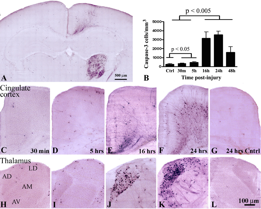
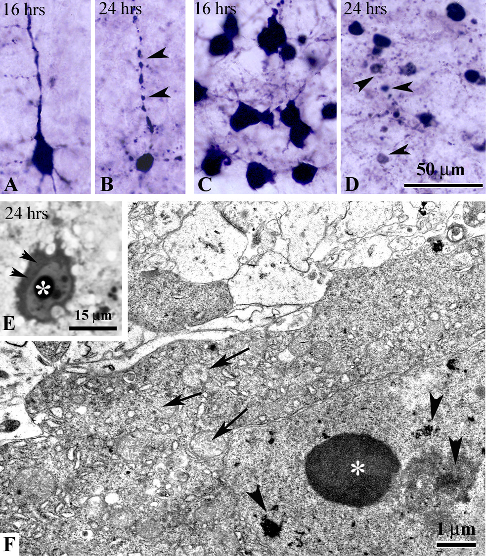
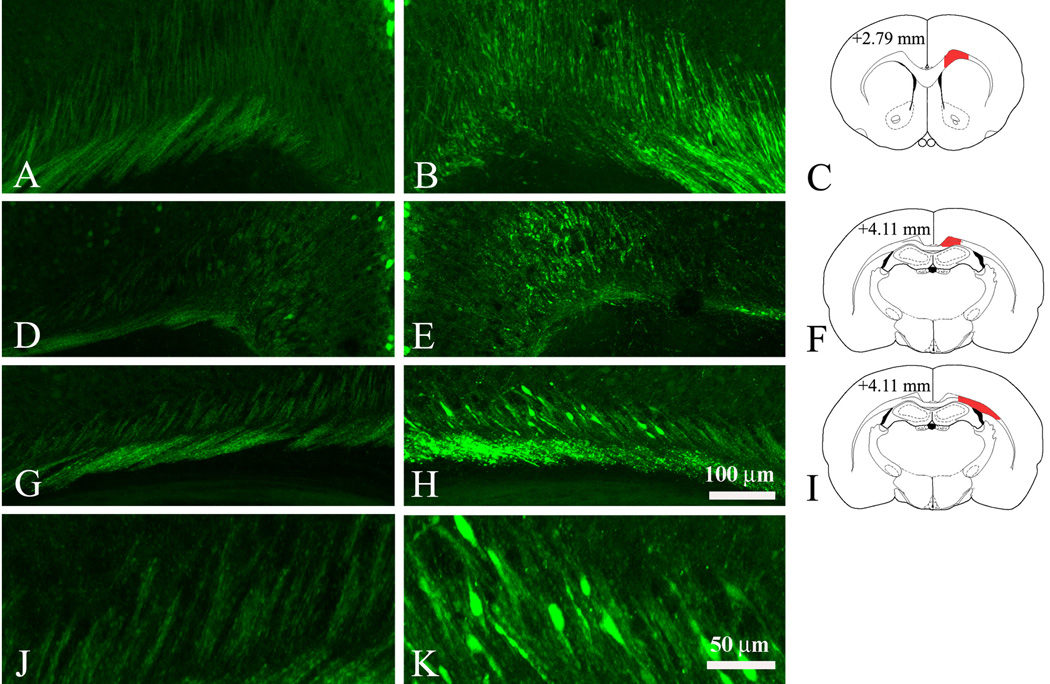
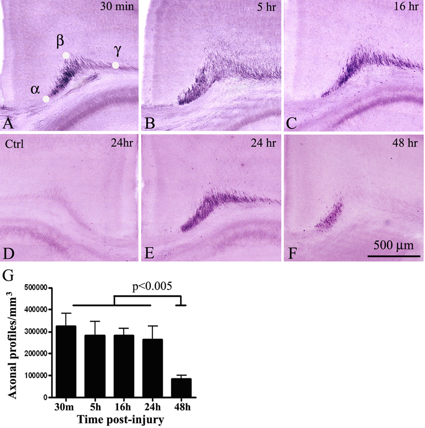
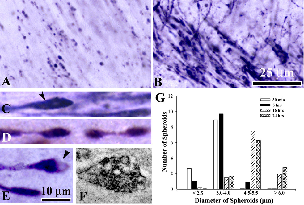
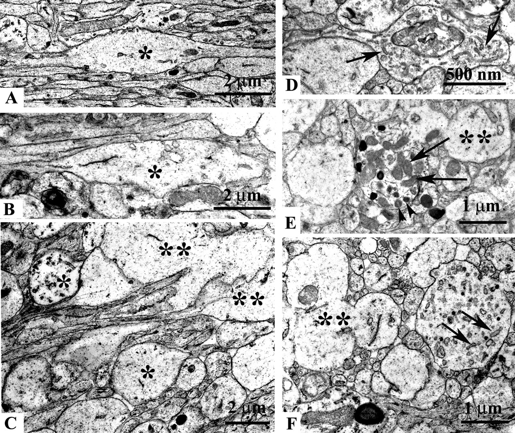
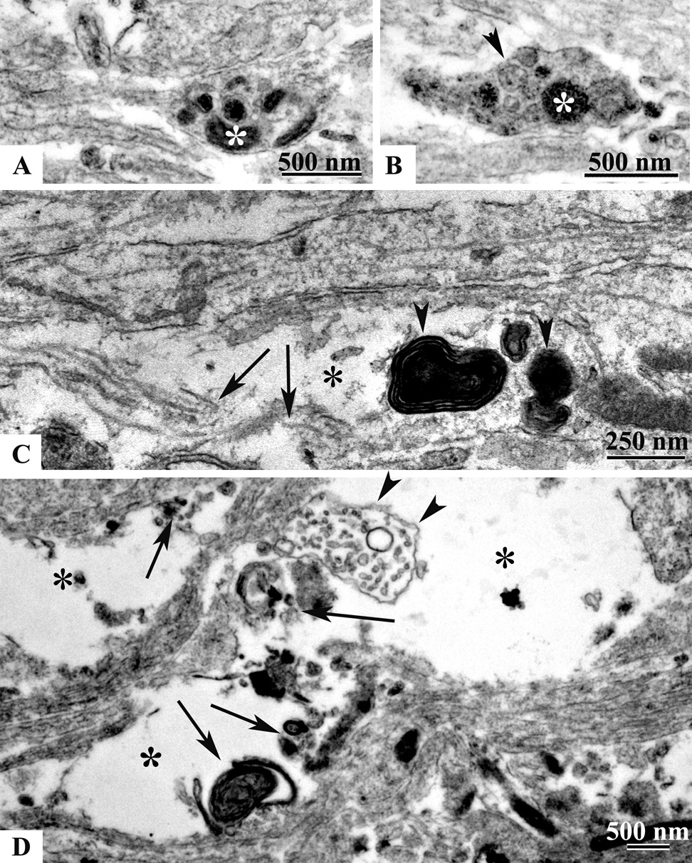
The immature brain in the first several years of childhood is very vulnerable to trauma. Traumatic brain injury (TBI) during this critical period often leads to neuropathological and cognitive impairment. Previous experimental studies in rodent models of infant TBI were mostly concentrated on neuronal degeneration, while axonal injury and its relationship to cell death have attracted much less attention. To address this, we developed a closed controlled head injury model in infant (P7) mice and characterized the temporospatial pattern of axonal degeneration and neuronal cell death in the brain following mild injury. Using amyloid precursor protein (APP) as marker of axonal injury we found that mild head trauma causes robust axonal degeneration in the cingulum/external capsule as early as 30 min post-impact. These levels of axonal injury persisted throughout a 24 h period, but significantly declined by 48 h. During the first 24 h injured axons underwent significant and rapid pathomorphological changes. Initial small axonal swellings evolved into larger spheroids and club-like swellings indicating the early disconnection of axons. Ultrastructural analysis revealed compaction of organelles, axolemmal and cytoskeletal defects. Axonal degeneration was followed by profound apoptotic cell death in the posterior cingulate and retrosplenial cortex and anterior thalamus which peaked between 16 and 24 h post-injury. At early stages post-injury no evidence of excitotoxic neuronal death at the impact site was found. At 48 h apoptotic cell death was reduced and paralleled with the reduction in the number of APP-labeled axonal profiles. Our data suggest that early degenerative response to injury in axons of the cingulum and external capsule may cause disconnection between cortical and thalamic neurons, and lead to their delayed apoptotic death.


Figures

Fig.1
Fig.1
A – Low power image of a representative coronal section showing the pattern of caspase-3 immunoreactivity in the mouse brain 24 hours following mild TBI. Time course of caspase-3 activation in the posterior cingulate cortex (C–G) and anterior thalamus (H–L) at 30 min (C, H), 5 hrs (D, I), 16 hrs (E,J) and 24 hrs (F, K) after mild injury in P7 mice. No caspase-3 positive cells are detected in the contralateral side (G, L). Note that at 24 hrs post injury additional anterior thalamic nuclei, including the laterodorsa (LD), anteroventral (AM), and to a lesser extent, the anteromedial (AM) show robust caspase-3 immunoreactivity (K). B – Quantitaive analysis of apoptotic cell death progression in the anterodorsal (AD) nucleus of thalamus between 30 min and 48 hrs post-injury.
Fig.2
Fig.2
Morphological changes in degenerating apoptotic neurons in the cortex (A, B) and AD thalamus (C, D). At 16 hrs post-injury caspase-3 positive cortical (A) and thalamic (C) neurons show intense somatic as well as dendritic staining. At 24 hrs, cortical neurons become shrunken (B) with fragmented apical dendritic shafts (arrowheads in B). Many apoptotic bodies (arrowheads in D) and cellular debris are detected in AD thalamus. E – high power light micrograph of apoptotic neuron in the posterior cingulate cortex (plastic section) showing condensation of the cytoplasm (arrowheads) and chromatin balls in the nucleus (asterisk) at 24 hrs. F – electron micrograph of a degenerating neuron in the AD thalamus at 24 hrs post-injury with typical ultrastructural morphological features of apoptosis: condensed cytoplasm, swelling of mitochondria (arrows), intranuclear chromatin balls (asterisk) and fragmentation of the nucleolus (arrowheads).
Fig.3
Fig.3
Identification of subcortical white matter tracts which are affected by mild injury in Thy1-YFP16 mouse at 16 hrs after impact. Axonal injury is determined by the accumulation of very strong YFP-labeling in axonal fibers and swellings in the trauma side (B, E, H, K). Most axons in the contralateral side (A, D, G, J) show smooth YFP-labeling with occasionally appearing swellings. C, F, and I are diagrams of the rostro-caudal level presented in coronal YFP images; areas of interest are filled in red. In the cingulum and external capsule of the trauma side numerous YFP-labeled swollen axons are detected at levels +2.79 (B, C) and + 4.11 (E, F and H, I). J and K are high magnification images taken from G and H showing morphological detail and increased YFP fluorescence in axonal swellings at the trauma side (K) compared to the contralateral side (J). Coordinates show the distance (in mm) from the frontal pole and are adapted from the P6 mouse atlas (Paxinos, Halliday, Watson and Koucherov eds., “Atlas of the developing mouse”, Academic Press, 2007). Scale bar, 100 µm (A, B, D, E, G, H), 50 µm (J, K).
Fig.4
Fig.4
Time-course of axonal injury in the cingulum/external capsule in P7 mice after mild TBI. Strong APP staining is detected as early as 30 min post-injury (A), and persists at 5 (B), 16 (C) and 24 hrs (E) after trauma. A substantial reduction in APP-immunoreactivity is observed 48 hrs after injury (F). No APP-labeled axons are detected in the contralateral side at all time points (D- representative control section at 24 hrs). F – Quantitative analysis of the density of APP-positive axonal profiles in the cingulum/external capsule at different time-points post-trauma. No significant difference is found between 5, 16 and 24 hrs when compared to 30 min, while substantial reduction in the number of APP-profiles is detected 48 hrs post trauma. α,β,γ in (A) represent reference points determining the triangular area used for axonal profile counts.
Fig.5
Fig.5
Morphological changes in degenerating APP-immonoreactive axons in the cingulum/external capsule. A – at 30 min post-injury many, predominantly small round APP-stained spheroids are detected. At 5 hrs, numerous injured axons appear as long and thick immunoreactive fibers ending in larger spheroids and swellings (B). At 5 to 24 hrs following mild TBI morphological changes in degenerating axons include the formation of clubs (C, arrowhead), beaded varicose fibers (D), vacuolated spheroids or bulbs (E, arrowhead). Scale bar, 25 µm (A,B), 100 µm (C, D, E). F - electron micrograph showing APP-stained vesiculated spheroid. I – Diameter-frequency histogram demonstrating changes in axonal swelling size at different time-points post-injury: 30 min (white bar), 5hrs (black bar), 16 hrs (striped bar) and 24 hrs (cross-hatched bar).
Fig. 6
Fig. 6
Ultrastructural axonal changes in the cingulum and external capsule at 5 hrs after mild TBI. Electron micrograph showing local swelling of an injured non-myelinated axon (A, asterisk) and normal appearing axonal segments (A, the lower left side). Note, that the majority of axonal profiles in this micrograph are sectioned longitudinally. Some axonal segments end as clubs or spheroids (single asterisks in B and C), others show disrupted axolemmal membranes (double asterisks in C, E and F). Longitudinal (D) and cross sections (E and F) of swellings showing accumulation of elongated vesicles (arrows in D and F), dense-core vesicles (arrowheads in E) and mitochondria (arrows in E). All swollen segments are devoid of cytoskeletal elements.
Fig. 7
Fig. 7
Ultrastructural axonal changes in the cingulum and external capsule at 24 hrs after mild TBI. Accumulation of mitochondria (asterisk in A and B) and vesicles (arrowhead in B) in axonal spheroids at 24 hrs are similar to that seen at 5 hrs. However, in many axonal club-like swellings (C, asterisk) signs of advanced degeneration such as edema, microtubule discontinuity (C, arrows) and accumulation of myelin figures (C, arrowheads) are present. Large vacuous spaces in the cingulum/external capsule (D, asterisks), remnants of a swollen axonal profiles (D, arrowheads) and scattered axoplasmic debris (D, arrows) are also observed.

References

    1. Adelson PD, Robichaud P, Hamilton RL, Kochanek PM. A model of diffuse traumatic brain injury in the immature rat. J Neurosurg. 1996;85:877–884. - PubMed
    1. Adelson PD, Jenkins LW, Hamilton RL, Robichaud P, Tran MP, Kochanek PM. Histopathologic response of the immature rat to diffuse traumatic brain injury. J Neurotrauma. 2001;18:967–976. - PubMed
    1. Anderson V, Catroppa C, Morse S, Haritou F, Rosenfeld J. Functional plasticity or vulnerability after early brain injury? Pediatrics. 2005;116:1374–1382. - PubMed
    1. Bailey CD, Johnson GV. Developmental regulation of tissue transglutaminase in the mouse forebrain. J Neurochem. 2004;91:1369–1379. - PubMed
    1. Bayly PV, Black EE, Pedersen RC, Leister EP, Genin GM. In vivo imaging of rapid deformation and strain in an animal model of traumatic brain injury. J Biomech. 2006a;39:1086–1095. - PMC - PubMed

Publication types

MeSH terms