. 2008 Jul;295(1):L152-61.
doi: 10.1152/ajplung.00515.2007. Epub 2008 Apr 25.

Utility of magnetic resonance imaging and nuclear magnetic resonance-based metabolomics for quantification of inflammatory lung injury

Affiliations

Utility of magnetic resonance imaging and nuclear magnetic resonance-based metabolomics for quantification of inflammatory lung injury

Natalie J Serkova et al. Am J Physiol Lung Cell Mol Physiol. 2008 Jul.

Abstract

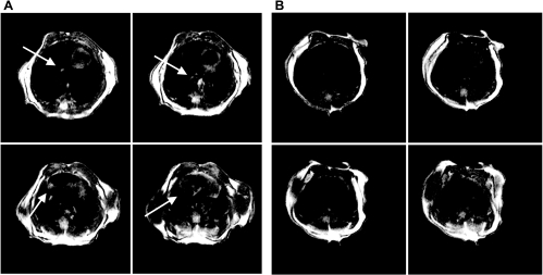
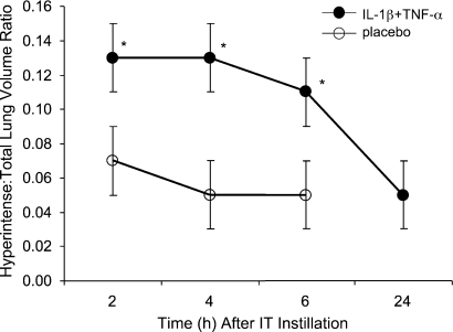
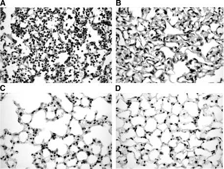
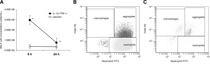
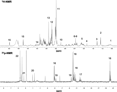
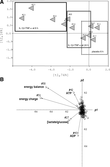
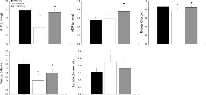
Magnetic resonance imaging (MRI) and metabolic nuclear magnetic resonance (NMR) spectroscopy are clinically available but have had little application in the quantification of experimental lung injury. There is a growing and unfulfilled need for predictive animal models that can improve our understanding of disease pathogenesis and therapeutic intervention. Integration of MRI and NMR could extend the application of experimental data into the clinical setting. This study investigated the ability of MRI and metabolic NMR to detect and quantify inflammation-mediated lung injury. Pulmonary inflammation was induced in male B6C3F1 mice by intratracheal administration of IL-1beta and TNF-alpha under isoflurane anesthesia. Mice underwent MRI at 2, 4, 6, and 24 h after dosing. At 6 and 24 h lungs were harvested for metabolic NMR analysis. Data acquired from IL-1beta+TNF-alpha-treated animals were compared with saline-treated control mice. The hyperintense-to-total lung volume (HTLV) ratio derived from MRI was higher in IL-1beta+TNF-alpha-treated mice compared with control at 2, 4, and 6 h but returned to control levels by 24 h. The ability of MRI to detect pulmonary inflammation was confirmed by the association between HTLV ratio and histological and pathological end points. Principal component analysis of NMR-detectable metabolites also showed a temporal pattern for which energy metabolism-based biomarkers were identified. These data demonstrate that both MRI and metabolic NMR have utility in the detection and quantification of inflammation-mediated lung injury. Integration of these clinically available techniques into experimental models of lung injury could improve the translation of basic science knowledge and information to the clinic.


Figures

Fig. 1.
Fig. 1.
Representative magnetic resonance imaging (MRI; 4 axial sections, 1 mm thick) of a mouse thorax that 6 h earlier received intratracheal (IT) interleukin (IL)-1β + tumor necrosis factor (TNF)-α (arrows indicate inflamed areas) (A) or received IT placebo (sterile saline) (B). Diffuse infiltrates are apparent in the images of the IL-1β+TNF-α-treated mouse. These changes corresponded to a hyperintense-to-total lung volume (HTLV) ratio of 0.15 compared with the placebo-treated mouse, for which the ratio was 0.04.
Fig. 2.
Fig. 2.
Time course of HTLV ratio for IL-1β+TNF-α- and placebo-treated mice. MRI was performed at 2, 4, and 6 h in the same mice. At these time points, the mean HTLV volume ratio was greater for IL-1β+TNF-α-treated mice than placebo-treated mice [*P = 0.04 (2 h), P = 0.001 (4 h), P = 0.03 (6 h)]. By 24 h, the mean (±SE) HTLV ratio of IL-1β+TNF-α-treated mice returned to control values (0.05 ± 0.00 and 0.07 ± 0.02, respectively; P = 0.52). Data are means (±SE) for 4 animals/group.
Fig. 3.
Fig. 3.
Representative light micrographs (×20) of hematoxylin and eosin-stained lung sections. A: 6 h after a mouse received IT IL-1β+TNF-α. The micrograph shows evidence of severe lung injury with an acute inflammatory response characterized by mononuclear inflammatory cell infiltrate and innumerable sloughed pneumocytes in the alveoli. The alveolar wall is edematous. Type II pneumocytes lining the alveoli are hypertrophic and hyperplastic. B: 6 h after a mouse received IT sterile saline (placebo). The micrograph shows empty alveolar spaces filled with air. The alveolar walls are lined with flattened epithelial cells. There are no inflammatory cells, no reactive type II pneumocytes, no fibrin, and no evidence of injury. C and D: 24 h after a mouse received either IT IL-1β+TNF-α (C) or IT sterile saline (D). Both micrographs show normal cellular morphology and no evidence of injury.
Fig. 4.
Fig. 4.
IT instillation of IL-1β+TNF-α resulted in an increase in bronchoalveolar lavage fluid (BALF) cell concentration (A) compared with placebo-treated mice at 6 h (P = 0.011). By 24 h the cell count had declined but was still greater than that in placebo-treated mice (P = 0.002). Data are means (±SE) of 4 animals/group. B and C: representative fluorescence-activated cell sorting (FACS) data showing the distribution (%) of leukocytes in BALF acquired from a single animal 6 h after IL-1β+TNF-α treatment (B) or placebo treatment (C). The mean (±SE) distribution of BALF neutrophils from all lavaged IL-1β+TNF-α-treated mice was 76.8 ± 2.2% compared with 1.6 ± 0.9% for all lavaged placebo-treated mice (P < 0.0001). Macrophages were less prevalent in IL-1β+TNF-α-treated mice than in placebo-treated mice (89.1 ± 2.0% vs. 0.6 ± 0.4%, P < 0.0001). This shift in the differential reflects the acute nature of the inflammation and is further evidenced by the presence of more aggregates, which most likely represent neutrophil-macrophage interactions, in the IL-1β+TNF-α-treated mice.
Fig. 5.
Fig. 5.
Representative 1H- and 31P-nuclear magnetic resonance (NMR) spectra of control lung tissue that provide quantitative information on 52 endogenous metabolites and ratios. Peak assignment of major metabolites on 1H- and 31P-NMR spectra: 1, valine, leucine, isoleucine; 2, lactate; 3, alanine; 4, acetate, 5, CH3-acetyl groups; 6–9, glutamate, succinate, glutamine, and total glutathione; 10, creatine; 11, total choline; 12, taurine; 13, glycine; 14, inositol, 15, glucose; 16, ATP; 17, sugar phosphates; 18, NAD+; 19, ADP; 20, phosphodiesters; 21, AMP; 22, phosphomonoesters.
Fig. 6.
Fig. 6.
Principal component analysis (PCA) scores on 52 quantitative metabolic end points of lung extracts. A: PCA scores (ti) based on the global metabolic pattern clusters control for IL-1β+TNF-α-treated and placebo-treated mice. Each triangle represents the quantitative metabolic data set from an individual animal for which 1–4 represent IL-1β+TNF-α-treated mice at 6 h, 5–8 represent IL-1β+TNF-α-treated mice at 24 h, and 9–11 represent placebo-treated mice at 6 h. B: PCA plots (pi) on individual data sets distinguish single putative biomarkers responsible for the clustering pattern observed in A. Each circle represents a specific metabolic end point (from the panel of 52 metabolites included in the PCA).
Fig. 7.
Fig. 7.
Quantitative comparison of distinguished metabolic biomarkers for IL-1β+TNF-α-treated mice, which shows the reversible nature of the metabolic derangement induced by the cytokines. The energy balance was calculated as ATP-to-ADP ratios and the energy charge as [(ATP + 0.5ADP)/(ATP + ADP + AMP)]. All measured indexes were different from control at 6 h (*P ≤ 0.03), with the exception of ADP. Differences were also evident between the 6 h and 24 h time points (+P < 0.05). By 24 h, ATP levels and the lactate-to-glucose ratio had recovered to control levels (P = 0.11 and P = 0.07, respectively). The amount of ADP increased over the study's time course, most likely because of injury-induced increase in energy production. Data are means ± SE of 3 or 4 animals/group.

References

    1. Ashcroft T, Simpson JM, Timbrell V. Simple method of estimating severity of pulmonary fibrosis on a numerical scale. J Clin Pathol 41: 467–470, 1988. - PMC - PubMed
    1. Beckmann N, Cannet C, Karmouty-Quintana H, Tigani B, Zurbruegg S, Ble FX, Cremillieux Y, Trifilieff A. Lung MRI for experimental drug research. Eur J Radiol 64: 381–396, 2007. - PubMed
    1. Beckmann N, Tigani B, Ekatodramis D, Borer R, Mazzoni L, Fozard JR. Pulmonary edema induced by allergen challenge in the rat: noninvasive assessment by magnetic resonance imaging. Magn Reson Med 45: 88–95, 2001. - PubMed
    1. Brealey D, Brand M, Hargreaves I, Heales S, Land J, Smolenski R, Davies NA, Cooper CE, Singer M. Association between mitochondrial dysfunction and severity and outcome of septic shock. Lancet 360: 219–223, 2002. - PubMed
    1. Caruthers SD, Paschal CB, Pou NA, Roselli RJ, Harris TR. Regional measurements of pulmonary edema by using magnetic resonance imaging. J Appl Physiol 84: 2143–2153, 1998. - PubMed

Publication types

Substances