

Superoxide dismutase protects against apoptosis and alveolar enlargement induced by ceramide
- PMID: 18441093
- PMCID: PMC2494787
- DOI: 10.1152/ajplung.00448.2007
Superoxide dismutase protects against apoptosis and alveolar enlargement induced by ceramide
Abstract
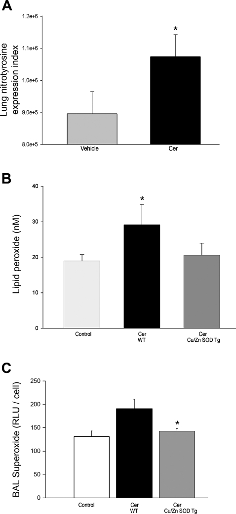
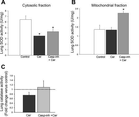
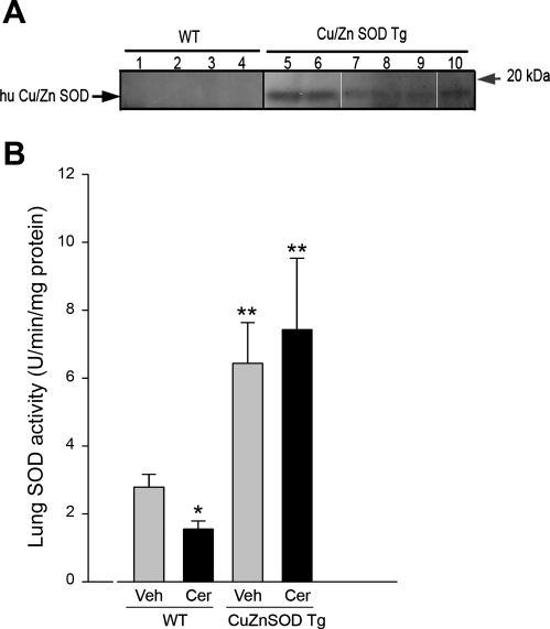
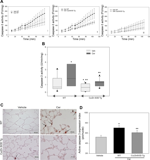
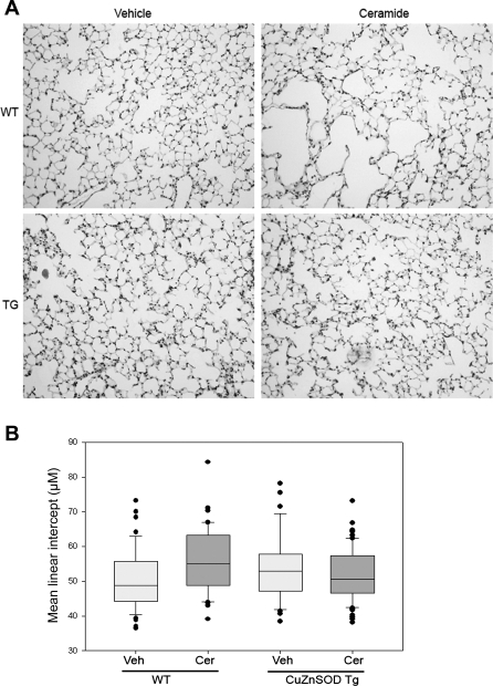
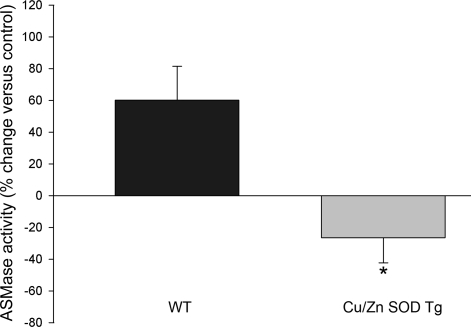
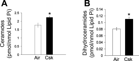
The molecular events leading to emphysema development include generation of oxidative stress and alveolar cell apoptosis. Oxidative stress upregulates ceramides, proapoptotic signaling sphingolipids that trigger further oxidative stress and alveolar space enlargement, as shown in an experimental model of emphysema due to VEGF blockade. As alveolar cell apoptosis and oxidative stress mutually interact to mediate alveolar destruction, we hypothesized that the oxidative stress generated by ceramide is required for its pathogenic effect on lung alveoli. To model the direct lung effects of ceramide, mice received ceramide intratracheally (Cer(12:0) or Cer(8:0); 1 mg/kg) or vehicle. Apoptosis was inhibited with a general caspase inhibitor. Ceramide augmentation shown to mimic levels found in human emphysema lungs increased oxidative stress, and decreased, independently of caspase activation, the lung superoxide dismutase activity at 48 h. In contrast to their wild-type littermates, transgenic mice overexpressing human Cu/Zn SOD were significantly protected from ceramide-induced superoxide production, apoptosis, and air space enlargement. Activation of lung acid sphingomyelinase in response to ceramide treatment was abolished in the Cu/Zn SOD transgenic mice. Since cigarette smoke-induced emphysema in mice is similarly ameliorated by the Cu/Zn SOD overexpression, we hypothesized that cigarette smoke may induce ceramides in the mouse lung. Utilizing tandem mass spectrometry, we documented increased lung ceramides in adult mice exposed to cigarette smoke for 4 wk. In conclusion, ceramide-induced superoxide accumulation in the lung may be a critical step in ceramide's proapoptotic effect in the lung. This work implicates excessive lung ceramides as amplifiers of lung injury through redox-dependent mechanisms.
Figures
Similar articles
-
RTP801 is required for ceramide-induced cell-specific death in the murine lung.Am J Respir Cell Mol Biol. 2013 Jan;48(1):87-93. doi: 10.1165/rcmb.2012-0254OC. Epub 2012 Sep 28. Am J Respir Cell Mol Biol. 2013. PMID: 23024063 Free PMC article.
-
Stimulation of sphingosine 1-phosphate signaling as an alveolar cell survival strategy in emphysema.Am J Respir Crit Care Med. 2010 Feb 15;181(4):344-52. doi: 10.1164/rccm.200906-0826OC. Epub 2009 Dec 3. Am J Respir Crit Care Med. 2010. PMID: 19965812 Free PMC article.
-
Oxidative stress and apoptosis interact and cause emphysema due to vascular endothelial growth factor receptor blockade.Am J Respir Cell Mol Biol. 2003 Jul;29(1):88-97. doi: 10.1165/rcmb.2002-0228OC. Epub 2003 Jan 31. Am J Respir Cell Mol Biol. 2003. PMID: 12600822
-
Role of apoptosis in the pathogenesis of COPD and pulmonary emphysema.Respir Res. 2006 Mar 30;7(1):53. doi: 10.1186/1465-9921-7-53. Respir Res. 2006. PMID: 16571143 Free PMC article. Review.
-
Lung cancer and lung injury: the dual role of ceramide.Handb Exp Pharmacol. 2013;(216):93-113. doi: 10.1007/978-3-7091-1511-4_5. Handb Exp Pharmacol. 2013. PMID: 23563653 Free PMC article. Review.
Cited by
-
Pathogenesis of chronic obstructive pulmonary disease.J Clin Invest. 2012 Aug;122(8):2749-55. doi: 10.1172/JCI60324. Epub 2012 Aug 1. J Clin Invest. 2012. PMID: 22850885 Free PMC article. Review.
-
Development and Challenges of Diclofenac-Based Novel Therapeutics: Targeting Cancer and Complex Diseases.Cancers (Basel). 2022 Sep 9;14(18):4385. doi: 10.3390/cancers14184385. Cancers (Basel). 2022. PMID: 36139546 Free PMC article. Review.
-
NLRP3/Caspase-1 inflammasome activation is decreased in alveolar macrophages in patients with lung cancer.PLoS One. 2018 Oct 26;13(10):e0205242. doi: 10.1371/journal.pone.0205242. eCollection 2018. PLoS One. 2018. PMID: 30365491 Free PMC article.
-
Subcutaneous administration of neutralizing antibodies to endothelial monocyte-activating protein II attenuates cigarette smoke-induced lung injury in mice.Am J Physiol Lung Cell Mol Physiol. 2019 Mar 1;316(3):L558-L566. doi: 10.1152/ajplung.00409.2018. Epub 2019 Jan 10. Am J Physiol Lung Cell Mol Physiol. 2019. PMID: 30628489 Free PMC article.
-
Neutral sphingomyelinase 2: a novel target in cigarette smoke-induced apoptosis and lung injury.Am J Respir Cell Mol Biol. 2011 Mar;44(3):350-60. doi: 10.1165/rcmb.2009-0422OC. Epub 2010 May 6. Am J Respir Cell Mol Biol. 2011. PMID: 20448054 Free PMC article.
References
-
- Aherne WA, Dunnill MS. Morphometry. London: Edward Arnold, 1982, p. 205.
-
- Andrieu-Abadie N, Gouaze V, Salvayre R, Levade T. Ceramide in apoptosis signaling: relationship with oxidative stress. Free Radic Biol Med 31: 717–728, 2001. - PubMed
-
- Bartalesi B, Cavarra E, Fineschi S, Lucattelli M, Lunghi B, Martorana PA, Lungarella G. Different lung responses to cigarette smoke in two strains of mice sensitive to oxidants. Eur Respir J 25: 15–22, 2005. - PubMed
-
- Borg J, London J. Copper/zinc superoxide dismutase overexpression promotes survival of cortical neurons exposed to neurotoxins in vitro. J Neurosci Res 70: 180–189, 2002. - PubMed
-
- Cantin A, Crystal RG. Oxidants, antioxidants and the pathogenesis of emphysema. Eur J Respir Dis Suppl 139: 7–17, 1985. - PubMed
Publication types
MeSH terms
Substances
Grants and funding
LinkOut - more resources
Full Text Sources
Other Literature Sources
Molecular Biology Databases
Research Materials
Miscellaneous