. 2008 Jun;294(6):H2814-21.
doi: 10.1152/ajpheart.00095.2008. Epub 2008 Apr 25.

Effect of ANG II on endothelial cell apoptosis and survival and its impact on skeletal muscle angiogenesis after electrical stimulation

Affiliations

Effect of ANG II on endothelial cell apoptosis and survival and its impact on skeletal muscle angiogenesis after electrical stimulation

Micheline M de Resende et al. Am J Physiol Heart Circ Physiol. 2008 Jun.

Abstract

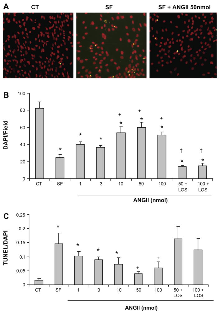
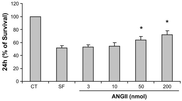
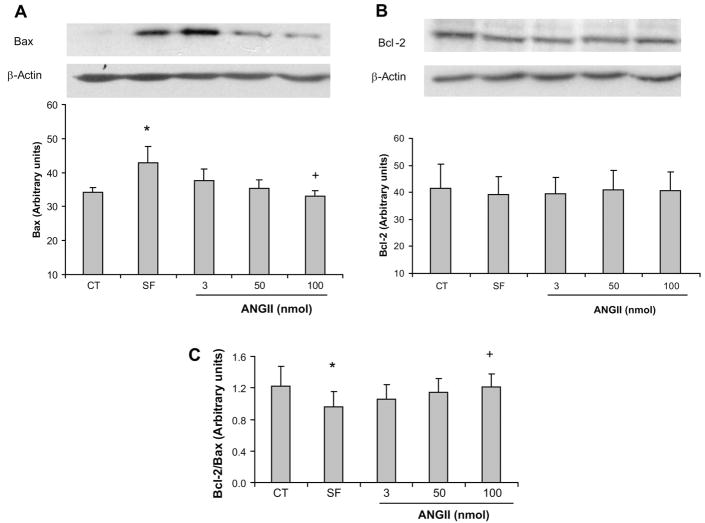
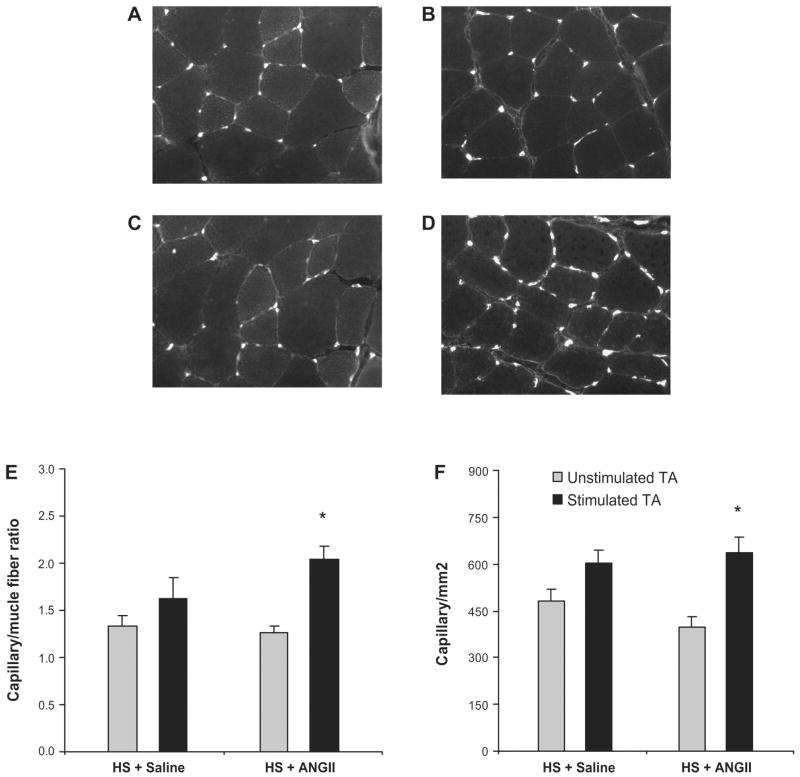
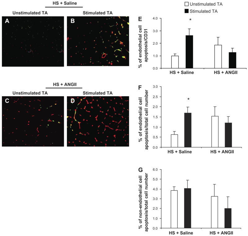
We have previously shown that skeletal muscle angiogenesis induced by electrical stimulation is significantly attenuated when SS-13BN/Mcwi rats are fed a high-salt diet. This effect was associated with a large increase in endothelial cell (EC) apoptosis. We hypothesized that the low levels of ANG II during high-salt diet would increase EC apoptosis and consequently diminish the angiogenic response. To test this hypothesis, a series of in vitro and in vivo studies was performed. EC apoptosis and viability were evaluated after incubation with ANG II under serum-free conditions. After 24 h of incubation, ANG II increased EC viability and Bcl-2-to-Bax ratio along with a dose-dependent decrease in EC apoptosis. This effect was blocked by the ANG II type 1 receptor antagonist losartan. To confirm our in vitro results, ANG II (3 ng.kg(-1).min(-1)) was chronically infused in rats fed a high-salt diet (4% NaCl). ANG II decreased EC apoptosis and produced a significant increase (40%) in skeletal muscle angiogenesis after electrical stimulation. These in vivo results were in agreement with our in vitro results and demonstrate that the attenuation of ANG II levels during a high-salt diet may induce EC apoptosis and consequently block the angiogenic response induced by electrical stimulation. Furthermore, under normal conditions, ANG II increases EC viability and protects EC from apoptosis possibly by inactivation of the mitochondrial apoptotic pathway.


Figures

Fig. 1
Fig. 1
ANG II blocks serum deprivation-induced apoptosis in microvascular endothelial cells (ECs) through the angiotensin type 1 (AT1) receptor. Cells were treated with either serum-free medium or serum free + ANG II (1–100 nmol/l). A: representative fluorescence images of terminal deoxynucleotidyl transferase-mediated dUTP nick end labeling (TUNEL) assays from cells in RPMI medium and 20% serum (CT), RPMI serum free (SF), and serum-free medium plus 50 nmol ANG II (SF + ANG II 50 nmol). Apoptotic cells are shown in yellow. Magnification, ×20. B: total number of cells stained by 4′,6-diamidino-2-phenylindole dihydrochloride (DAPI) after 24 h incubation period. C: dose-response analysis of the antiapoptotic effects of ANG II after 24 h incubation period. Values represent means ± SE. Each point is the mean of data from duplicate experiments. *P < 0.05 vs. CT; +P < 0.05 vs. SF; †P < 0.05 vs. respective ANG II dose.
Fig. 2
Fig. 2
Dose-dependent effects of ANG II on EC survival. Cell viability in complete RPMI medium (20% serum) was considered to be 100%. EC viability was determined after 24 h incubation in serum-free medium or ANG II (3, 10, 50, and 200 nmol). Error bars represent SDs of 5 independent experiments performed in triplicate. *P < 0.05 vs. SF.
Fig. 3
Fig. 3
A and B, top: effect of ANG II on Bax and Bcl-2 expression in ECs in culture. AC, bottom: Bcl-2-to-Bax ratio. Values represent means ± SE. P < 0.05 vs. control (*) and vs. SF (+); n = 5 experiments.
Fig. 4
Fig. 4
Effect of ANG II on capillary density after 7 days of electrical stimulation. A and B: representative images of cross sections of the tibialis anterior (TA) muscle showing the capillaries and fibers in the unstimulated and stimulated TA muscle of SS-13BN/Mcwi rats fed a high-salt (HS) diet that received saline infusion, respectively. C and D: representative images of cross sections of the TA muscle showing capillaries and fibers in the unstimulated and stimulated TA muscle of SS-13BN/Mcwi rats fed a high-salt diet that received ANG II infusion, respectively. E: quantitative capillary-to-fiber ratio analysis. F: capillary/mm2; n = 8 in the HS + saline group and n = 7 in the HS + ANG II group. Values are expressed as means ± SE. *P < 0.05 vs. unstimulated TA.
Fig. 5
Fig. 5
ANG II acts as a survival factor in skeletal muscle vascular ECs of SS-13BN fed a high salt diet. A–D: double immunofluorescent staining to detect apoptotic ECs in the TA muscle of SS-13BN rats fed a high-salt diet. A and B: representative images of unstimulated and stimulated TA of rats fed a high-salt diet and infused with saline, respectively. Red: ECs; green: apoptotic cells; yellow: apoptotic ECs. C and D: representative images of unstimulated and stimulated TA of rats fed a high-salt diet and infused with ANG II, respectively. E: percentage of EC apoptosis as a function of the total number of ECs. F: percentage of EC apoptosis as a function of the total number of cells. G: percentage of non-EC apoptosis as a function of the total number of cells. Values are expressed as means ± SE. *P < 0.05 vs. unstimulated TA; n = 9 in the HS + saline group and n = 6 in the HS + ANG II group. Values represent means ± SE. *P < 0.05 vs. unstimulated TA.

Similar articles

Cited by

References

    1. Amaral SL, Linderman JR, Greene AS. Angiogenesis induced by electrical stimulation is mediated by angiotensin II. Microcirculation. 2000;8:57–67. - PubMed
    1. Amaral SL, Papanek P, Greene AS. Angiotensin II and VEGF are involved in angiogenesis induced by short term exercise training. Am J Physiol Heart Circ Physiol. 2001;281:H1163–H1169. - PubMed
    1. Amaral SL, Roman RJ, Greene AS. Renin Gene transfer restores angiogenesis and vascular endothelial growth factor expression in Dahl-S rats. Hypertension. 2001;37:386–390. - PubMed
    1. Amaya K, Ohta T, Kitagawa H, Kayahara M, Takamura H, Fujimura T, Nishimura G, Shimizu K, Miwa K. Angiotensin II activates MAP kinase and NF-kappaB through angiotensin II type I receptor in human pancreatic cancer cells. Int J Oncol. 2004;25:849–856. - PubMed
    1. Arafat HA, Gong Q, Chipitsyna G, Rizvi A, Saa CT, Yeo CJ. Anti-hypertensives as novel antineoplastics: angiotensin-I-converting enzyme inhibitors and angiotensin II type 1 receptor blockers in pancreatic ductal adenocarcinoma. J Am Coll Surg. 2007;204:996–1005. - PubMed

Publication types

MeSH terms