. 2008 Apr;20(4):1073-87.
doi: 10.1105/tpc.107.055632. Epub 2008 Apr 25.

Functional and physiological characterization of Arabidopsis INOSITOL TRANSPORTER1, a novel tonoplast-localized transporter for myo-inositol

Affiliations

Functional and physiological characterization of Arabidopsis INOSITOL TRANSPORTER1, a novel tonoplast-localized transporter for myo-inositol

Sabine Schneider et al. Plant Cell. 2008 Apr.

Abstract

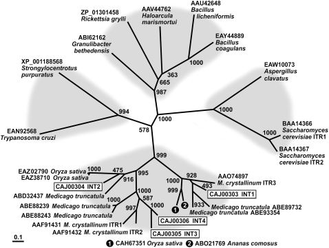
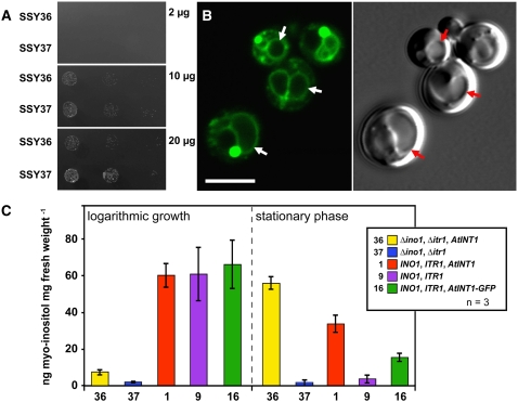
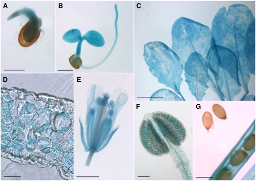
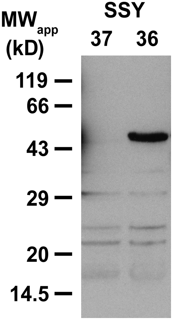
Arabidopsis thaliana INOSITOL TRANSPORTER1 (INT1) is a member of a small gene family with only three more genes (INT2 to INT4). INT2 and INT4 were shown to encode plasma membrane-localized transporters for different inositol epimers, and INT3 was characterized as a pseudogene. Here, we present the functional and physiological characterization of the INT1 protein, analyses of the tissue-specific expression of the INT1 gene, and analyses of phenotypic differences observed between wild-type plants and mutant lines carrying the int1.1 and int1.2 alleles. INT1 is a ubiquitously expressed gene, and Arabidopsis lines with T-DNA insertions in INT1 showed increased intracellular myo-inositol concentrations and reduced root growth. In Arabidopsis, tobacco (Nicotiana tabacum), and Saccharomyces cerevisiae, fusions of the green fluorescent protein to the C terminus of INT1 were targeted to the tonoplast membranes. Finally, patch-clamp analyses were performed on vacuoles from wild-type plants and from both int1 mutant lines to study the transport properties of INT1 at the tonoplast. In summary, the presented molecular, physiological, and functional studies demonstrate that INT1 is a tonoplast-localized H(+)/inositol symporter that mediates the efflux of inositol that is generated during the degradation of inositol-containing compounds in the vacuolar lumen.


Figures

Figure 1.
Figure 1.
Phylogenetic Tree of the INT Family of Arabidopsis Inositol Transporters and Related Transport Proteins from Other Species Using Maximum Likelihood. The phylogenetic tree was calculated from confirmed or predicted inositol transporter sequences. The tree reveals four different clusters: metazoa and protozoa on the left, fungi on the right, bacteria and archaea at the top, and plants, with two clearly distinguishable subgroups, at the bottom. Bootstrap samples of 1000 samplings are shown at each branch. GenBank accession numbers and names of the organisms are given plus, where available, the published name of the protein. Arabidopsis INT proteins are boxed.
Figure 2.
Figure 2.
Characterization of the Δitr1 Δino1 Yeast Double Mutant Expressing the INT1 cDNA in the Sense (SSY36) or Antisense (SSY37) Orientation. (A) Growth of SSY36 and SSY37 on minimal media supplemented with the indicated concentrations of myo-inositol. (B) Subcellular localization of INT1-GFP in SSY16 cells. The images show yeast cells with one or more vacuoles that were photographed either under GFP excitation light (confocal section; left) or under white light (Nomarski section; right). Vacuoles of some cells are marked with arrows. Bar = 5 μm. (C) Ion chromatographic analyses of myo-inositol concentrations determined in whole-cell ethanol extracts of SSY1 (1), SSY9 (9), SSY16 (16), SSY36 (36), and SSY37 (37) cells that were grown in liquid medium with 10 μg/mL myo-inositol and harvested either during the logarithmic growth phase or in the stationary phase. The genotypes of the different yeast strains are shown in the inset.
Figure 3.
Figure 3.
Subcellular Localization of INT1-GFP in the Tonoplast Membranes of Arabidopsis Protoplasts or of Tobacco Epidermis Cells. (A) Confocal section of an intact Arabidopsis protoplast expressing the INT1-GFP fusion construct. The arrow shows the tonoplast membrane passing a chloroplast on the inner side. (B) Projection of 40 confocal sections taken from an Arabidopsis protoplast expressing the INT1-GFP fusion construct. (C) Confocal section of a hatched Arabidopsis vacuole after lysis of the plasma membrane (fluorescence and white light images merged). (D) Confocal section of a tobacco epidermis cell bombarded with the INT1-GFP construct. The arrow marks the tonoplast membrane at the inner side of a chloroplast (enlarged in the inset). Red fluorescence in (A), (B), and (D) shows chlorophyll autofluorescence. Bars = 8 μm in (A) to (D) and 2 μm in the inset of (D).
Figure 4.
Figure 4.
Analyses of GUS Histochemical Staining in INT1 Promoter/GUS Plants. (A) and (B) Germinating (A) and young (B) seedling with GUS staining in the cotyledons, the hypocotyl, and the root. (C) Rosette leaves showing uneven, cloudy GUS staining. (D) Cross section of a rosette leaf with GUS staining in the mesophyll. (E) Fully developed flower with staining in all floral organs. (F) Closed anther with weak GUS staining in all cells and stronger staining in the pollen grains. (G) Silique with almost mature seeds. No GUS staining is detected in the seeds, but it is detected in all other tissues of the silique. Bars = 50 μm in (D), 100 μm in (F), 0.5 mm in (A), (B), and (G), 1 mm in (E), and 5 mm in (C).
Figure 5.
Figure 5.
Protein Gel Blot of Total Membrane Proteins from SSY36 and SSY37 Yeast Cells. Identification of recombinant INT1 protein with affinity-purified αINT1 (dilution, 1:10) in SDS extracts from yeast total membranes (10 μg/lane) after separation on a polyacrylamide gel and transfer to nitrocellulose.
Figure 6.
Figure 6.
Characterization of int1.1 and int1.2 Mutant Plants on the Genomic and mRNA Levels. (A) Schematic presentation of the INT1 gene (white boxes, exons; black lines, introns; hatched areas, 5′ and 3′ flanking regions). Positions of T-DNA insertions, left borders (LB), and binding sites for the primers (arrows) used in (B) are indicated. (B) PCR analyses of genomic int1.1 and int1.2 DNA. Heterozygous and homozygous plants were discriminated by PCR performed with wild-type-specific or mutant (m)–specific pairs of primers (int1.1 plants, int1/679f and int1/1749r for the wild type, LBb1 and int1-2/1749r for the mutant; int1.2 plants, int1/-945 and int1/557r for the wild type, LBb1 and int1/557r for the mutant). (C) Mutant analyses on the mRNA level. Fragments were amplified from wild-type and mutant cDNAs that corresponded to the full INT1 mRNA or to mRNA fragments upstream or downstream from the T-DNA insertions. Control reactions were performed with primers for the Arabidopsis ACT2 cDNA.
Figure 7.
Figure 7.
Phenotypic Characterization of int1.1 and int1.2 Mutant Plants. (A) Comparison of the root growth of int1.1 and wild-type seedlings (14 d old) on synthetic medium supplemented with the indicated concentrations of myo-inositol (mg/L). (B) Quantitative analysis of root lengths of int1.1 (yellow) and wild-type (red) seedlings (10 d old) on synthetic medium supplemented with the indicated concentrations of myo-inositol (n = 12; ±sd). (C) Comparison of myo-inositol, glucose, fructose, and sucrose concentrations in ethanol extracts from rosette leaves of int1.1 plants (red), of wild-type plants (yellow), and of int2.1 (blue) and int4.2 (green) mutant plants (n = 9; ±sd) that were grown, harvested, and analyzed in parallel. (D) A similar data set for extracts (n = 10) of independently grown and analyzed int1.2 (red) and wild-type (yellow) plants. The significance of the difference between the inositol concentrations in int1.2 and wild-type extracts was tested with Student's t test (P = 4 × 10⁻⁶).
Figure 8.
Figure 8.
Whole-Vacuole Recordings of Currents Triggered by myo-Inositol in Mesophyll Vacuoles of INT1 Wild-Type and int1 Mutant Plants. (A) The stylized presentation of vacuoles with attached patch pipettes shows the experimental conditions in the absence (gray bars) or presence (black bars) of myo-inositol in the bath medium. Cytosolic application of 50 mM myo-inositol (black bars) resulted in INT1-mediated H+ currents into the vacuoles of wild-type plants, as represented by the upward deflection of the current traces. Under the same experimental conditions, no or significantly smaller myo-inositol–induced H+ currents were observed in int1.1 or int1.2 mutant plants, respectively. Note that INT1-dependent currents were determined in the inverse mode (i.e., pH values in the extravacuolar bath and the patch pipette solution were adjusted to 5.5 and 7.5, respectively). (B) Similar measurement as in (A), but with 50 mM glucose on vacuoles from wild-type and int1.1 mutant plants. (C) Average currents calculated from several experiments (n = 3; ±sd) performed under identical conditions as the original recordings shown in (A) and (B). Reproducibly, inositol induced no current (= 0 pA/pF) in vacuoles of int1.1 plants.

Similar articles

Cited by

References

    1. Abel, S., and Theologis, A. (1994). Transient transformation of Arabidopsis leaf protoplasts: A versatile experimental system to study gene expression. Plant J. 5 421–427. - PubMed
    1. Alonso, J.M., et al. (2003). Genome-wide insertional mutagenesis of Arabidopsis thaliana. Science 301 653–657. - PubMed
    1. Aluri, S., and Büttner, M. (2007). Identification and functional expression of the Arabidopsis thaliana vacuolar glucose transporter 1 and its role in seed germination and flowering. Proc. Natl. Acad. Sci. USA 104 2537–2542. - PMC - PubMed
    1. Barth, I., Meyer, S., and Sauer, N. (2003). PmSUC3: Characterization of a SUT2/SUC3-type sucrose transporter from Plantago major. Plant Cell 15 1375–1385. - PMC - PubMed
    1. Becker, D., Kemper, E., Schell, J., and Masterson, R. (1992). New plant binary vectors with selectable markers located proximal to the left T-DNA border. Plant Mol. Biol. 20 8123–8128. - PubMed

Publication types

Associated data