. 2008 Jun 27;283(26):18066-75.
doi: 10.1074/jbc.M801213200. Epub 2008 Apr 25.

Direct binding of integrin alphavbeta3 to FGF1 plays a role in FGF1 signaling

Affiliations

Direct binding of integrin alphavbeta3 to FGF1 plays a role in FGF1 signaling

Seiji Mori et al. J Biol Chem. .

Abstract

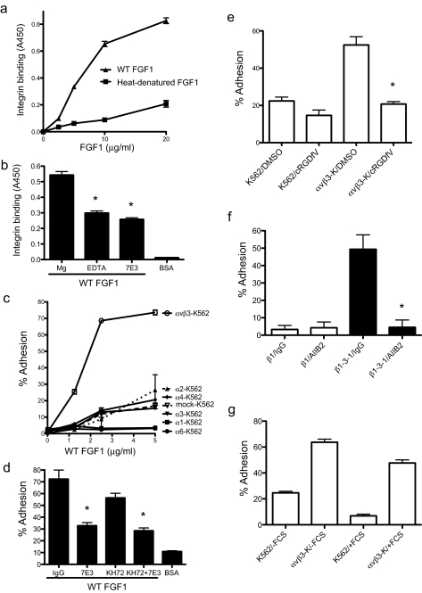
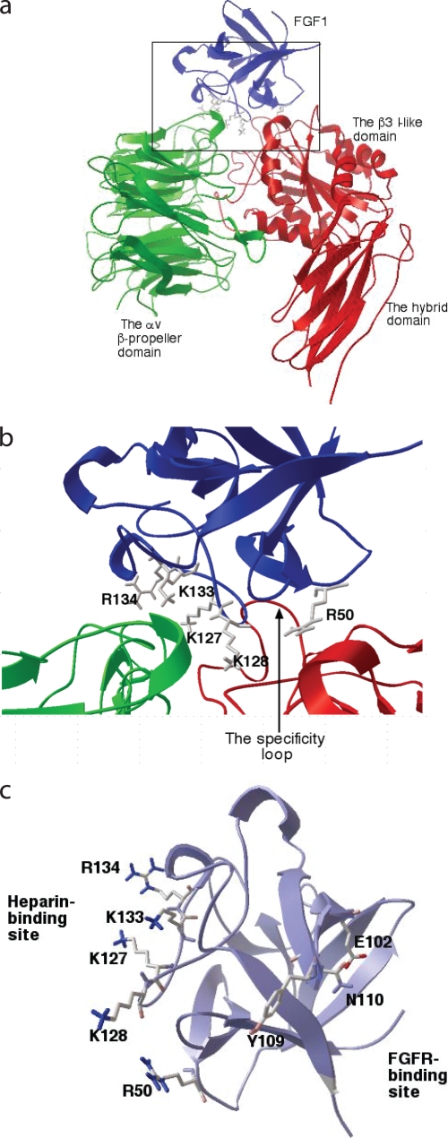
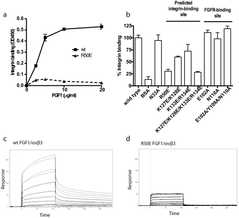
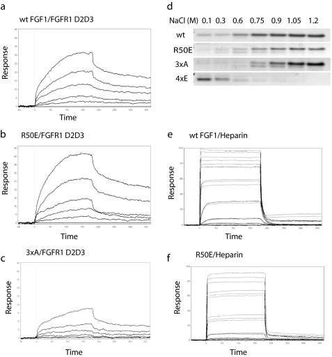
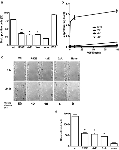
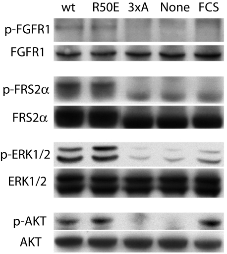
Integrins play a role in fibroblast growth factor (FGF) signaling through cross-talk with FGF receptors (FGFRs), but the mechanism underlying the cross-talk is unknown. We discovered that FGF1 directly bound to soluble and cell-surface integrin alphavbeta3 (K(D) about 1 microm). Antagonists to alphavbeta3 (monoclonal antibody 7E3 and cyclic RGDfV) blocked this interaction. alphavbeta3 was the predominant, if not the only, integrin that bound to FGF1, because FGF1 bound only weakly to several beta1 integrins tested. We presented evidence that the CYDMKTTC sequence (the specificity loop) within the ligand-binding site of beta3 plays a role in FGF1 binding. We found that the integrin-binding site of FGF1 overlaps with the heparin-binding site but is distinct from the FGFR-binding site using docking simulation and mutagenesis. We identified an FGF1 mutant (R50E) that was defective in integrin binding but still bound to heparin and FGFR. R50E was defective in inducing DNA synthesis, cell proliferation, cell migration, and chemotaxis, suggesting that the direct integrin binding to FGF1 is critical for FGF signaling. Nevertheless, R50E induced phosphorylation of FGFR1 and FRS2alpha and activation of AKT and ERK1/2. These results suggest that the defect in R50E in FGF signaling is not in the initial activation of FGF signaling pathway components, but in the later steps in FGF signaling. We propose that R50E is a useful tool to identify the role of integrins in FGF signaling.


Figures

FIGURE 1.
FIGURE 1.
Direct binding of FGF1 to integrin αvβ3. a, recombinant soluble αvβ3 bound to WT FGF1 but not to heat-denatured FGF1. Wells of 96-well microtiter plates were coated with WT or heat-denatured FGF1 at the indicated concentrations, and the remaining protein-binding sites were blocked with BSA. Recombinant soluble αvβ3 was added to the wells and incubated for 1 h at room temperature. Bound αvβ3 was determined by using HRP-conjugated anti-His6 antibody and peroxidase substrate at A450. Data are shown as means ± S.E. of triplicate experiments. b, effects of antibodies and EDTA on the binding of soluble αvβ3 to FGF1. The ELISA-type integrin-binding assay was performed as described above. Soluble αvβ3 was incubated with 10 μg/ml 7E3 or control mouse IgG for 10 min on ice prior to adding to the wells. Concentrations of MgCl2 and EDTA are 10 and 5 mm, respectively. BSA was used as a negative control. Data are shown as means ± S.E. of triplicate experiments. *, p < 0.05, n = 3 by t test. c, adhesion of K562 cells that express different integrins to FGF1. Wells of 96-well microtiter plates were coated with FGF1, and the remaining protein-binding sites were blocked with BSA. Cells were added to the wells and incubated for 1 h at 37 °C in Hepes-Tyrode's buffer containing 1 mm MnCl2, and bound cells were quantified after removing unbound cells by using phosphatase assays. Data are shown as means ± S.E. of triplicate experiments. d, inhibition of cell adhesion to FGF1 by anti-integrin antibodies. Adhesion assay was performed as described in c. Cells were incubated with 10 μg/ml control mouse IgG, mAb 7E3 (anti-β3, function blocking), or KH72 (anti-α5) for 30 min on ice prior to adding to the wells. Data are shown as means ± S.E. of triplicate experiments. *, p < 0.05 to control IgG, n = 3 by t test. e, inhibition of αvβ3-FGF1 interaction by cyclic RGDfV, a specific antagonist to αvβ3. Adhesion assay was performed as described in c. Cyclic RGDfV was used at 10 μm, and DMSO, in which stock cyclic RGDfV was solubilized, was used as a control. *, p = 0.0024 betweenαvβ3-K562 (αvβ3-K)/DMSO andαvβ3-K562/cyclic RGDfV. f, binding of theβ1–3-1 mutant to FGF1. Adhesion assay was performed as described in c. Cells were incubated with 10 μg/ml control mouse IgG or mAb AIIB2 (anti-β1, function blocking). Data are shown as means ± S.E. of triplicate experiments. *, p < 0.05 to DMSO, n = 3 by t test. g, effect of FCS to αvβ3-FGF1 interaction. Adhesion assay was performed as described in c. Assays were performed in the absence and presence of FCS (10%). Data are shown as means ± S.E. of triplicate experiments.
FIGURE 2.
FIGURE 2.
Identification of the integrin-binding site in FGF1. a, model of FGF1-integrin interaction. Docking simulation of the interaction between FGF1 (PDB code 1AXM) and integrin αvβ3 (PDB code 1L5G) was performed as described under the “Experimental Procedures” using AutoDock3. Model a of 1AXM was used for docking. The headpiece of 1L5G was used as a receptor. The pose in the cluster 1 with the lowest docking energy -26.3 kcal/mol is shown. This pose represents the most stable pose of FGF1 when FGF1 interacts with integrin αvβ3. b, positions of amino acid residues that are selected for mutagenesis at the predicted interface between FGF1 and αvβ3. Several amino acid residues within the predicted integrin-binding site in FGF1 were selected for mutagenesis. c, positions of the amino acid residues (E102A, Y109A, and N110A) at the FGFR-binding site selected for mutagenesis. Note that the predicted integrin-binding site is distinct from the FGFR-binding site.
FIGURE 3.
FIGURE 3.
Effect of FGF mutations on the binding to integrin αvβ3. a, R50E mutation of FGF1 reduced its binding to integrinαvβ3. Amino acid residues in the integrin-binding site or in the FGFR-binding site were mutated individually or in groups. WT FGF1 and the R50E mutant were coated to a plastic plate at various concentrations as indicated, and the binding of soluble αvβ3 was determined as described in Fig. 1a. The results show that the R50E mutation reduced αvβ3 binding. b, summary of the effects of FGF1 mutations on integrin binding. The results suggest that mutations in the predicted integrin-binding site of FGF1 reduced the binding of solubleαvβ3 to FGF1, but mutations in the FGFR-binding site did not. c and d, binding of WT FGF1 (c) and R50E FGF1 (d) to αvβ3 in SPR. Soluble αvβ3 was immobilized on a CM5 sensor chip. WT and R50E FGF1 were individually 2-fold serially diluted from 8 μm to 125 nm in HBS-P buffer with 1 mm of Mn2+, and the 3xA FGF1 was 2-fold serially diluted from 6 μm to 93.75 nm in the same buffer. 4xE did not show any binding to integrin (data not shown).
FIGURE 4.
FIGURE 4.
Effect of FGF mutations on the binding to FGFR and heparin. a–c, binding of WT (a), R50E (b), and 3xA (c) FGF1 mutant to FGFR1 D2D3 fragment in SPR. FGFR1 D2D3 was immobilized to a CM5 sensor chip. WT or mutant FGF1 was 2-fold diluted serially from 800 to 50 nm. KD is calculated as 30 pm for WT FGF1, 14 pm for R50E, 5.5 μm for 4xE, and 0.41 mm for 3xA. d, binding of R50E to heparin-Sepharose. We incubated partially purified WT and mutant FGF1 with heparin-Sepharose and eluted with increasing concentrations of NaCl. Eluted proteins were analyzed by SDS-PAGE and proteins stained with Coomassie Brilliant Blue. e and f, binding of WT FGF1 (e) and R50E FGF1 (f) to heparin in SPR. Biotinylated heparin was immobilized to a streptavidin sensor chip. 5-fold serially diluted WT, R50E, and 3xA ranging from 800 nm to 51.2 pm, and 2-fold serially diluted 4xE ranging from 1.6 μm to 50 nm in HBS-EP were injected at 50 μl/min for 3 min. KD is calculated as 16.6 nm for WT FGF1, 66 nm for R50E, and 11.5 nm for 3xA. 4xE did not show any binding to heparin (data not shown).
FIGURE 5.
FIGURE 5.
Integrin-binding defective FGF1 mutants are defective in FGF signaling. a, DNA synthesis. Balb 3T3 cells were plated on coverslips in 6-well culture plates, serum-starved for 48 h, and stimulated with 5 ng/ml WT or mutant FGF1s in the presence of 5 μg/ml heparin for 24 h. BrdUrd was added to the medium for the last 6 h of the incubation. BrdUrd-positive cells were counted. Results are shown as means ± S.E. of triplicate experiments. *, the BrdUrd incorporation was lower in cells treated with R50E (p = 0.0003), 4xE (p = 0.019), and 3xA (p = 0.003) than with WT FGF1. We used 2-way analysis of variance for statistical analysis. Similar results were obtained using NIH3T3 cells. FCS, 10% FCS was added to the medium as a positive control. b, proliferation of BaF3 cells that express human FGFR1c (BaF3-FR1c). BaF3-FR1c cells were maintained for 48 h with WT or mutant FGF1 at indicated concentrations instead of IL-3, and cell proliferation was measured by MTS assays. Results are shown as means ± S.E. of triplicate experiments. c, in vitro scratch wound healing. Confluent serum-starved Balb 3T3 cells were scratched. After washing with serum-free medium, the cells were incubated in DMEM containing 5 μg/ml heparin and 5 ng/ml WT or mutant FGF1s for 24 h at 37 °C. The rate of wound healing was quantified by using ImageJ. d, chemotaxis. The bottom of the polycarbonate filter of the Transwell apparatus was coated with fibronectin. The lower chamber contained serum-free DMEM containing 5 ng/ml WT or the mutants of FGF1. Balb 3T3 cells (105 cells/filter) were plated on filter and incubated 37 °C for 24 h, and the cells were stained with crystal violet. The cells that migrated to the bottom side of the membrane were counted. Results are expressed as means of the number of chemotaxed cells in three fields. *, the number of chemotaxed cells was lower with R50E (p = 0.026), 4xE (p = 0.0004), and 3xA (p = 0.0078) than with WT FGF1. We used 2-way analysis of variance for statistical analysis.
FIGURE 6.
FIGURE 6.
Effect of FGF1 mutations on FGF signaling. FCS, 10% FCS. Serum-starved NIH3T3 cells were stimulated with WT or mutant FGF1s (5 ng/ml) in the presence of 5 μg/ml heparin for 10 min at 37 °C. For FGFR phosphorylation (p-FGFR1), phosphorylated FGFR1 was first immunoprecipitated from lysates (750 μl, total 2.6 mg of protein) using anti-phospho-Tyr antibodies and then blotted using anti-FGFR1 antibodies. Whole FGFR1 in cell lysates were detected with anti-FGFR1. For FRS2α, ERK1/2, and AKT, cell lysates were analyzed by Western blotting with antibodies against phospho-FRS2α, FRS2α, phospho-ERK1/2, ERK1/2, phospho-AKT, and AKT, respectively. Results with the 4xE mutant were very similar to those with the 3xA mutant (data not shown).

References

    1. Presta, M., Dell'Era, P., Mitola, S., Moroni, E., Ronca, R., and Rusnati, M. (2005) Cytokine Growth Factor Rev. 16 159-178 - PubMed
    1. Ullrich, A., and Schlessinger, J. (1990) Cell 61 203-212 - PubMed
    1. Powers, C. J., McLeskey, S. W., and Wellstein, A. (2000) Endocr.-Relat. Cancer 7 165-197 - PubMed
    1. Klint, P., and Claesson-Welsh, L. (1999) Front. Biosci. 4 D165-D177 - PubMed
    1. Thisse, B., and Thisse, C. (2005) Dev. Biol. 287 390-402 - PubMed

Publication types

Substances