. 2008 Jun 27;283(26):18402-10.
doi: 10.1074/jbc.M709819200. Epub 2008 Apr 25.

Entry to "formula tunnel" revealed by SLC4A4 human mutation and structural model

Affiliations

Entry to "formula tunnel" revealed by SLC4A4 human mutation and structural model

Min-Hwang Chang et al. J Biol Chem. .

Abstract

Glaucoma, cataracts, and proximal renal tubular acidosis are diseases caused by point mutations in the human electrogenic Na(+) bicarbonate cotransporter (NBCe1/SLC4A4) (1, 2). One such mutation, R298S, is located in the cytoplasmic N-terminal domain of NBCe1 and has only moderate (75%) function. As SLC transporters have high similarity in their membrane and N-terminal primary sequences, we homology-modeled NBCe1 onto the crystal structure coordinates of Band 3(AE1) (3). Arg-298 is predicted to be located in a solvent-inaccessible subsurface pocket and to associate with Glu-91 or Glu-295 via H-bonding and charge-charge interactions. We perturbed these putative interactions between Glu-91 and Arg-298 by site-directed mutagenesis and used expression in Xenopus oocyte to test our structural model. Mutagenesis of either residue resulted in reduced transport function. Function was "repaired" by charge reversal (E91R/R298E), implying that these two residues are interchangeable and interdependent. These results contrast the current understanding of the AE1 N terminus as protein-binding sites and propose that hkNBCe1 (and other SLC4) cytoplasmic N termini play roles in controlling HCO(3)(-) permeation.


Figures
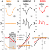
FIGURE 1.
FIGURE 1.
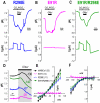
Voltage-clamped hkNBCe1 physiology. A-C, water-injected oocytes (A), oocytes expressing wild-type hkNBCe1 (B), and R298S mutants (C) were voltage-clamped at -60 mV, and passing currents I(μAmp) and pHi were measured simultaneously. Data shown are representative experimental traces from different clones, and the sample size of each clone is indicated in Table 1. D, expanded time scale of Na+ removal from panel (gray box) over 6.6 min before and after Na+ was removed from the formula image solution. NBCe1 transport formula image into the cell working against acidification due to the CO2 exposure resulting in alkalized pHi before the Na+ removal was only observed in WT-hkNBCe1. After Na+ removal, pHi change (pH units/s) of WT-hkNBCe1 (-130 × 10-5 pH units/s) decreased fastest among the three (R298S = -71, × 10-5; water = -10 × 10-5). E and F, current-voltage relationship of hkNBCe1 mutants. Oocytes were voltage-clamped at -60 mV and stepped as indicated under “Experimental Procedures.” Data shown are corrected I-V traces by subtracting initial measurements in ND-96 at corresponding voltage-steps; sample sizes are in parentheses. E, averaged data collected in CO2/formula image when the elicited outward currents reached a steady state. The reversal potentials are -80 mV. F, averaged traces when the inward current reached a steady state in the 0Na+-CO2/formula image solution. The WT and R298S currents all rectified at positive holding voltages with no obvious reversal potential.
FIGURE 2.
FIGURE 2.
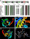
Structural model of the N-terminal cytoplasmic domain (62-313) of hkNBCe1. A, sequence alignment of hkNBCe1 amino acid sequences 81-92, 101-114, 209-218, 225-235, and 293-306 with corresponding regions of other SLC4 bicarbonate transporter proteins. The intensity of the shading corresponds to the consensus level of the conserved residues in the gene family. The asterisk indicates missense human mutation R298S. Residues marked as green are residues within 4 Å from Glu-91, Arg-295, and Arg-298 (high-lighted in red) and highly likely to have charge interactions with these three residues. B, ribbon diagram structure of hkNBCe1 amino acid sequence 62-313 mapped onto the corresponding region of the Band 3 (human AE1) crystal structure (PDB number 1HYN) of Low and co-workers (3). The blue to red progressive color scheme denotes secondary structure succession from N terminus toward C terminus. C, human missense mutation R298S is located in the solvent-inaccessible pocket. Highlighted are residues Glu-91 and Glu-295 about 3.5 Å from the Arg-298 side chains putatively forming H-bond (green) with Arg-298. Side-chain charges of the three residues are color-coded to negative (red) and positive (blue). D, chain of polar residues creating a polar channel in the domain core. E, close up view on the hydrogen bond network of polar residues. The NCBI/GenBank™ accession numbers for these sequences are hkNBCe1 (human kidney form electrogenic Na+ bicarbonate cotransporter 1; AF007216), hAE1-hAE3 (human anion exchanger 1-3; M27819, U62531, and U05596), drNDAE1 (Drosophila sodium-dependent anion exchanger 1; AF047468), ceNBC (Caenorhabditis elegans Na+ bicarbonate cotransporter; AF004926), aNBCe1 (Ambystoma electrogenic Na+ bicarbonate cotransporter 1; AF001958), rkNBCe1 (rat kidney electrogenic Na+ bicarbonate cotransporter 1; NM_053424), mNBCe1 (murine electrogenic Na+ bicarbonate cotransporter 1; AF141934), rb1NBCe1 and -2 (rat brain electrogenic Na+ bicarbonate cotransporter 1 and 2; AF124441 and AF254802), hpNBCe1 (human pancreas form electrogenic Na+ bicarbonate cotransporter 1; AF053753), NBCe1-dace (Osorezan dace electrogenic Na+ bicarbonate cotransporter 1; AB055467), NBCe1-trout (rainbow trout Na+ bicarbonate cotransporter 1; AF434166), NBC3 (human Na+ bicarbonate cotransporter 3; AF069512), NBC4c (human Na+ bicarbonate cotransporter 4; AF293337), NBCn1-D (rat electroneutral Na+ bicarbonate cotransporter 1-D, NM_058211), NCBE (Na+-Cl-/bicarbonate exchanger; AB040457), NDCBE(Na+-driven chloride/bicarbonate exchanger; AY151155).
FIGURE 3.
FIGURE 3.
Voltage-clamped hkNBCe1 mutants physiology. Oocytes expressing R298E (A), E91R (B), or E91R/R298E (C) mutants were voltage-clamped at -60 mV, and passing currents I(μAmp) and pHi were measured simultaneously as in Fig. 1. Data shown are representative experimental traces from different clones, and the sample size of each clone is indicated in Table 1. The R298E mutant has decreased currents and less substantial pHi changes in response to the addition of CO2/formula image and/or Na+ removal. The E91R mutant transport function is almost abolished in terms of current magnitude and pHi changes. Double mutant E91R/R298E operates approximately like a wild-type hkNBCe1. D, expanded time scale of Na+ removal from panel (gray box) over 6.6 min before and after Na+ was removed from the formula image solution. NBCe1 transports formula image into the cell, working against acidification due to CO2 exposure, resulting in increased pHi of different magnitudes prior to 0Na+. After Na+ removal, the pHi changes (×10-5 pH units/s) are E91R/R298E ≈ WT > R298E (= R298S) > E91R. E and F, current-voltage relationship of hkNBCe1 mutants. Data shown are corrected I-V traces by subtracting initial measurements in ND-96 at corresponding voltage-steps; numbers are in parentheses. E, averaged data collected in CO2/formula image. The reversal potentials are approximately -80 mV. F, averaged steady state inward current with 0Na+-CO2/formula image solution. The wild-type and mutants currents all rectified at positive holding voltages with no obvious reversal potential.
FIGURE 4.
FIGURE 4.
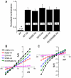
NBCe1 surface expression in oocytes. A, the normalized luminescence value (mean ± S. E.) of oocytes expressing HA-tagged hkNBCe1 mutants. The surface expression of the transporter protein on the oocytes was labeled by a monoclonal rat-α-HA 1° antibody and a goat-α-rat horseradish peroxidase-IgG 2° antibody and measured with a luminometer after adding chemiluminescent substrate. The luminescence values of the clones were normalized to the value of the WT-hkNBCe1 with HA tag. The oocytes were from three different donor frogs and sample sizes of each clone are shown in the bars. The asterisk indicates a luminescence value for that clone that is statistically different (p < 0.01) from that of HA-tagged WT-hkNBCe1. B and C, the current-voltage relationship of hkNBCe1 mutants in CO2/formula image (B) and in 0Na+-CO2/formula image solutions (C).
FIGURE 5.
FIGURE 5.
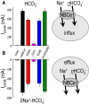
Summary of structural effects on hkNBCe1 function. A, averaged peak outward currents collected in CO2/formula image (pH 7.5) solution, i.e. inward NaHCO3 uptake (right). B, average peak inward current in 0Na+-CO2/formula image (pH 7.5) solution, i.e. outward renal transport (right). Oocytes expressing different hkNBC1 mutants and water-injected oocytes were voltage-clamped at -60 mV. The current changes were recorded in various experimental solutions, and peak current changes were calculated by subtracting the elicited currents before and after the formula image or 0Na+ formula image solutions were introduced. The sample size for each clone is in Table 1. R298S and R298E are mutants with less pronounced peak current changes, which indicates altered transport functions. E91R is a severe mutation with nearly abolished transport function having significantly less current responses to solution changes. R298S, E91R, and R298E currents are stoically different from WT and E91R/R298E. E91R/R298E double mutant has peak current changes identical to WT-hkNBCe1, indicating that E91R/R298E restores E91R to a fully WT-hkNBCe1-like function (not statistically different from WT).

Similar articles

Cited by

References

    1. Igarashi, T., Inatomi, J., Sekine, T., Cha, S. H., Kanai, Y., Kunimi, M., Tsukamoto, K., Satoh, H., Shimadzu, M., Tozawa, F., Mori, T., Shiobara, M., Seki, G., and Endou, H. (1999) Nat. Genet. 23 264-266 - PubMed
    1. Romero, M. F., and Smith, B. L. (2002) FASEB J. 16 a52-a53
    1. Zhang, D., Kiyatkin, A., Bolin, J. T., and Low, P. S. (2000) Blood 96 2925-2933 - PubMed
    1. Alper, S. L. (2002) Annu. Rev. Physiol. 64 899-923 - PubMed
    1. Romero, M. F. (2005) Curr. Opin. Nephrol. Hypertens. 14 495-501 - PubMed

Publication types