Review
. 2008 Apr 28;10(1):18.
doi: 10.1186/1532-429X-10-18.

Myocardial first-pass perfusion cardiovascular magnetic resonance: history, theory, and current state of the art

Affiliations
Review

Myocardial first-pass perfusion cardiovascular magnetic resonance: history, theory, and current state of the art

Bernhard L Gerber et al. J Cardiovasc Magn Reson. .

Abstract

In less than two decades, first-pass perfusion cardiovascular magnetic resonance (CMR) has undergone a wide range of changes with the development and availability of improved hardware, software, and contrast agents, in concert with a better understanding of the mechanisms of contrast enhancement. The following review provides a perspective of the historical development of first-pass CMR, the developments in pulse sequence design and contrast agents, the relevant animal models used in early preclinical studies, the mechanism of artifacts, the differences between 1.5T and 3T scanning, and the relevant clinical applications and protocols. This comprehensive overview includes a summary of the past clinical performance of first-pass perfusion CMR and current clinical applications using state-of-the-art methodologies.


Figures

Figure 1
Figure 1
Typical first-pass perfusion pulse sequence. (A) Acquisitions are cardiac gated using ECG signals. Multiple slices are acquired consecutively (shown: 6 slices every 2 R-R intervals). This is repeated continuously during the first-pass and washout of the contrast agent. (B) T1 contrast is generated using a saturation pulse (dark gray) followed by fast imaging of each slice (light gray) using gradient echo, gradient echo-planar, or steady state free precession acquisitions. Image contrast is primarily determined by the saturation recovery time, labeled TSR.
Figure 2
Figure 2
Comparison between a dark rim artifact (DRA, top) and a real perfusion defect (bottom) from two different patients. From left to right are shown the contrast arrival to the left ventricle, the myocardium, and finally recirculation. A DRA artifact usually lasts for a few heartbeats while a real defect tends to be more persistent.
Figure 3
Figure 3
Simulation of a short axis line profile. Comparison between a high and a low contrast (between the LV blood and the myocardium) line profile. Signal oscillations are higher for the high contrast image, as expected from the Gibbs truncation theory. (See Additional file 1.)
Figure 4
Figure 4
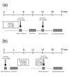
Typical protocol for assessing extent of microvascular obstruction with rest first-pass perfusion imaging. An alternate method of imaging microvascular obstruction is single shot late gadolinium enhancement.
Figure 5
Figure 5
(A) Typical first-pass perfusion protocol for adenosine stress perfusion CMR. (B) Typical first- pass perfusion protocol for dipyridamole stress perfusion imaging.
Figure 6
Figure 6
Myocardial perfusion CMR was performed in a 43 year-old female to evaluate exertional chest pressure. Perfusion images obtained during intravenous infusion of adenosine (A) demonstrate severe hypoenhancement of the septum, anterior wall, and apex that is not present on resting perfusion imaging (B). Late gadolinium enhancement acquisitions are negative for hyperenhancement (C); together, these findings suggest severe myocardial ischemia in the distribution of the left anterior descending coronary artery without infarction. (D) Invasive coronary angiography confirms high-grade serial stenoses in the left anterior descending coronary artery. (See Additional file 2.)
Figure 7
Figure 7
First-pass myocardial perfusion CMR was performed in a 75 year-old male with a history of bypass surgery seeking a second opinion regarding intervention for bypass graft disease. Perfusion images obtained during intravenous infusion of adenosine (A) demonstrates diffuse subendocardial perfusion abnormality not present on resting perfusion imaging (B). Late gadolinium enhancement acquisitions (C) show a small region of enhancement involving the lateral wall. Together, these findings suggested prior infarct with ischemia. His symptoms were controlled with optimization of anti-anginal medical therapies. (See Additional file 3.)

Similar articles

Cited by

References

    1. Hesse B, Tagil K, Cuocolo A, Anagnostopoulos C, Bardies M, Bax J, Bengel F, Busemann Sokole E, Davies G, Dondi M, Edenbrandt L, Franken P, Kjaer A, Knuuti J, Lassmann M, Ljungberg M, Marcassa C, Marie PY, McKiddie F, O'Connor M, Prvulovich E, Underwood R, van Eck-Smit B. EANM/ESC procedural guidelines for myocardial perfusion imaging in nuclear cardiology. Eur J Nucl Med Mol Imaging. 2005;32(7):855–897. doi: 10.1007/s00259-005-1779-y. - DOI - PubMed
    1. Sabharwal NK, Lahiri A. Role of myocardial perfusion imaging for risk stratification in suspected or known coronary artery disease. Heart. 2003;89(11):1291–1297. doi: 10.1136/heart.89.11.1291. - DOI - PMC - PubMed
    1. Zoghbi GJ, Iskandrian AE. Coronary artery disease: pharmacological stress. In: Zaret BL, Beller GA, editor. Nuclear Cardiology: State of the Art and Future Directions. 3. Philadelphia, PA: Mosby; 2005. pp. 233–253.
    1. Hachamovitch R, Berman DS, Shaw LJ, Kiat H, Cohen I, Cabico JA, Friedman J, Diamond GA. Incremental prognostic value of myocardial perfusion single photon emission computed tomography for the prediction of cardiac death: differential stratification for risk of cardiac death and myocardial infarction. Circulation. 97(6):535–543. Feb 17 1998. - PubMed
    1. Atkinson DJ, Burstein D, Edelman RR. First-pass cardiac perfusion: evaluation with ultrafast MR imaging. Radiology. 1990;174(3 Pt 1):757–762. - PubMed

MeSH terms