

Vascular mechanism of axonal degeneration in peripheral nerves in hemiplegic sides after cerebral hemorrhage: An experimental study
- PMID: 18442382
- PMCID: PMC2396626
- DOI: 10.1186/1749-7221-3-13
Vascular mechanism of axonal degeneration in peripheral nerves in hemiplegic sides after cerebral hemorrhage: An experimental study
Abstract
Background: Though retrograde neuronal death and vascular insufficiency have been well established in plegics following intracerebral hemorrhage, the effects of plegia on arterial nervorums of peripheral nerves have not been reported. In this study, the histopathological effects of the intracerebral hemorrhage on the dorsal root ganglions and sciatic nerves via affecting the arterial nervorums were investigated.
Methods: This study was conducted on 13 male hybrid rabbits. Three animals were taken as control group and did not undergo surgery. The remaining 10 subjects were anesthetized and were injected with 0.50 ml of autologous blood into their right sensory-motor region. All rabbits were followed-up for two months and then sacrificed. Endothelial cell numbers and volume values were estimated a three dimensionally created standardized arterial nervorums model of lumbar 3. Neuron numbers of dorsal root ganglions, and axon numbers in the lumbar 3 nerve root and volume values of arterial nervorums were examined histopathologically. The results were analyzed by using a Mann-Whitney-U test.
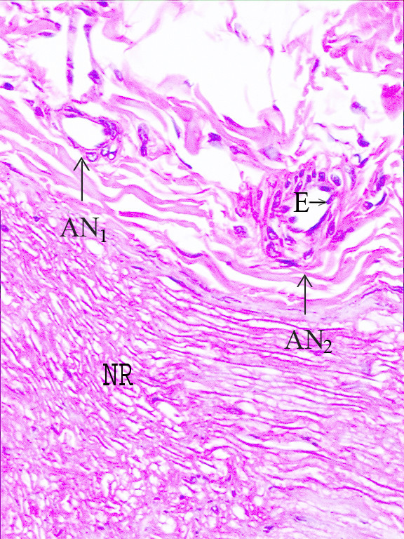
Results: Left hemiplegia developed in 8 animals. On the hemiplegic side, degenerative vascular changes and volume reduction in the arterial nervorums of the sciatic nerves, neuronal injury in the dorsal root ganglions, and axonal injury in the lumbar 3 were detected. Statistical analyses showed a significant correlation between the normal or nonplegic sides and plegic sides in terms of the neurodegeneration in the dorsal root ganglions (p < 0.005), axonal degeneration in the lumbar 3 nerve roots (p < 0.005), endothelial cell degeneration in the arterial nervorums (p < 0.001), and volume reduction in the arterial nervorums (p < 0.001).
Conclusion: Intracerebral hemorrhage resulted in neurodegeneration in the dorsal root ganglion and axonolysis in the sciatic nerves, endothelial injury, and volume reduction of the arterial nervorums in the sciatic nerves. The interruption of the neural network connection in the walls of the arterial nervorums in the sciatic nerves may be responsible for circulation disorders of the arterial nervorums, and arterial nervorums degeneration could result in sciatic nerves injury.
Figures


Similar articles
-
Unpublished Neuropathologic Mechanism Behind the Muscle Weakness/Paralysis and Gait Disturbances Induced by Sciatic Nerve Degeneration After Spinal Subarachnoid Hemorrhage: An Experimental Study.World Neurosurg. 2018 Nov;119:e1029-e1034. doi: 10.1016/j.wneu.2018.08.054. Epub 2018 Aug 23. World Neurosurg. 2018. PMID: 30144617
-
Ganglionary mechanisms of spasticity and ileus in cerebral hemorrhage: an experimental study.Int J Dev Neurosci. 2006 Nov;24(7):455-9. doi: 10.1016/j.ijdevneu.2006.07.001. Epub 2006 Sep 8. Int J Dev Neurosci. 2006. PMID: 16963220
-
The role of vagal nerve root injury on respiration disturbances in subarachnoid hemorrhage.Turk Neurosurg. 2015;25(2):273-8. doi: 10.5137/1019-5149.JTN.9964-13.1. Turk Neurosurg. 2015. PMID: 26014012
-
[Novel findings from an animal tourniquet shock model].Nihon Hoigaku Zasshi. 2003 Sep;57(2):125-34. Nihon Hoigaku Zasshi. 2003. PMID: 14574964 Review. Japanese.
-
Experience with examination of the spinal cord and peripheral nervous system (PNS) in mice: A brief overview.Exp Toxicol Pathol. 2014 Sep;66(7):277-80. doi: 10.1016/j.etp.2014.04.005. Epub 2014 May 24. Exp Toxicol Pathol. 2014. PMID: 24867273 Review.
Cited by
-
A Review of the Mysterious Roles of the Autonomic Ganglia Considered as Deep Intelligence Agency in the United States of the Brain.Eurasian J Med. 2023 Dec;55(1):S43-S48. doi: 10.5152/eurasianjmed.2023.23284. Eurasian J Med. 2023. PMID: 39128057 Free PMC article.
-
The impact of L5 dorsal root ganglion degeneration and Adamkiewicz artery vasospasm on descending colon dilatation following spinal subarachnoid hemorrhage: An experimental study; first report.J Craniovertebr Junction Spine. 2015 Apr-Jun;6(2):69-75. doi: 10.4103/0974-8237.156056. J Craniovertebr Junction Spine. 2015. PMID: 25972712 Free PMC article.
References
LinkOut - more resources
Full Text Sources
Other Literature Sources