

The abundant extrachromosomal DNA content of the Spiroplasma citri GII3-3X genome
- PMID: 18442384
- PMCID: PMC2386487
- DOI: 10.1186/1471-2164-9-195
The abundant extrachromosomal DNA content of the Spiroplasma citri GII3-3X genome
Abstract
Background: Spiroplama citri, the causal agent of citrus stubborn disease, is a bacterium of the class Mollicutes and is transmitted by phloem-feeding leafhopper vectors. In order to characterize candidate genes potentially involved in spiroplasma transmission and pathogenicity, the genome of S. citri strain GII3-3X is currently being deciphered.
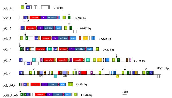
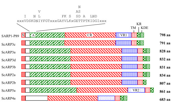
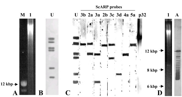
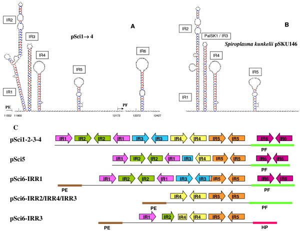
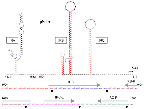
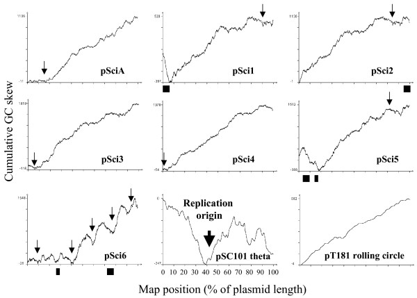
Results: Assembling 20,000 sequencing reads generated seven circular contigs, none of which fit the 1.8 Mb chromosome map or carried chromosomal markers. These contigs correspond to seven plasmids: pSci1 to pSci6, with sizes ranging from 12.9 to 35.3 kbp and pSciA of 7.8 kbp. Plasmids pSci were detected as multiple copies in strain GII3-3X. Plasmid copy numbers of pSci1-6, as deduced from sequencing coverage, were estimated at 10 to 14 copies per spiroplasma cell, representing 1.6 Mb of extrachromosomal DNA. Genes encoding proteins of the TrsE-TraE, Mob, TraD-TraG, and Soj-ParA protein families were predicted in most of the pSci sequences, in addition to members of 14 protein families of unknown function. Plasmid pSci6 encodes protein P32, a marker of insect transmissibility. Plasmids pSci1-5 code for eight different S. citri adhesion-related proteins (ScARPs) that are homologous to the previously described protein P89 and the S. kunkelii SkARP1. Conserved signal peptides and C-terminal transmembrane alpha helices were predicted in all ScARPs. The predicted surface-exposed N-terminal region possesses the following elements: (i) 6 to 8 repeats of 39 to 42 amino acids each (sarpin repeats), (ii) a central conserved region of 330 amino acids followed by (iii) a more variable domain of about 110 amino acids. The C-terminus, predicted to be cytoplasmic, consists of a 27 amino acid stretch enriched in arginine and lysine (KR) and an optional 23 amino acid stretch enriched in lysine, aspartate and glutamate (KDE). Plasmids pSci mainly present a linear increase of cumulative GC skew except in regions presenting conserved hairpin structures.
Conclusion: The genome of S. citri GII3-3X is characterized by abundant extrachromosomal elements. The pSci plasmids could not only be vertically inherited but also horizontally transmitted, as they encode proteins usually involved in DNA element partitioning and cell to cell DNA transfer. Because plasmids pSci1-5 encode surface proteins of the ScARP family and pSci6 was recently shown to confer insect transmissibility, diversity and abundance of S. citri plasmids may essentially aid the rapid adaptation of S. citri to more efficient transmission by different insect vectors and to various plant hosts.
Figures
Similar articles
-
Characterizing the replication and stability regions of Spiroplasma citri plasmids identifies a novel replication protein and expands the genetic toolbox for plant-pathogenic spiroplasmas.Microbiology (Reading). 2008 Oct;154(Pt 10):3232-3244. doi: 10.1099/mic.0.2008/019562-0. Microbiology (Reading). 2008. PMID: 18832328
-
Genome analysis of Spiroplasma citri strains from different host plants and its leafhopper vectors.BMC Genomics. 2021 May 22;22(1):373. doi: 10.1186/s12864-021-07637-8. BMC Genomics. 2021. PMID: 34022804 Free PMC article.
-
Sequence comparisons of plasmids pBJS-O of Spiroplasma citri and pSKU146 of S. kunkelii: implications for plasmid evolution.BMC Genomics. 2005 Dec 7;6:175. doi: 10.1186/1471-2164-6-175. BMC Genomics. 2005. PMID: 16336638 Free PMC article.
-
Spiroplasma plasmids.Isr J Med Sci. 1987 Jun;23(6):678-82. Isr J Med Sci. 1987. PMID: 3312107 Review.
-
Spiroplasmas: infectious agents of plants, arthropods and vertebrates.Wien Klin Wochenschr. 1997 Aug 8;109(14-15):604-12. Wien Klin Wochenschr. 1997. PMID: 9286068 Review.
Cited by
-
Deciphering Clostridium tyrobutyricum Metabolism Based on the Whole-Genome Sequence and Proteome Analyses.mBio. 2016 Jun 14;7(3):e00743-16. doi: 10.1128/mBio.00743-16. mBio. 2016. PMID: 27302759 Free PMC article.
-
Functional genomics of a Spiroplasma associated with the carmine cochineals Dactylopius coccus and Dactylopius opuntiae.BMC Genomics. 2021 Apr 6;22(1):240. doi: 10.1186/s12864-021-07540-2. BMC Genomics. 2021. PMID: 33823812 Free PMC article.
-
Sequences essential for transmission of Spiroplasma citri by its leafhopper vector, Circulifer haematoceps, revealed by plasmid curing and replacement based on incompatibility.Appl Environ Microbiol. 2010 May;76(10):3198-205. doi: 10.1128/AEM.00181-10. Epub 2010 Mar 19. Appl Environ Microbiol. 2010. PMID: 20305023 Free PMC article.
-
The enemy within: phloem-limited pathogens.Mol Plant Pathol. 2018 Jan;19(1):238-254. doi: 10.1111/mpp.12526. Epub 2017 Mar 9. Mol Plant Pathol. 2018. PMID: 27997761 Free PMC article. Review.
-
Genome sequence of the mesophilic Thermotogales bacterium Mesotoga prima MesG1.Ag.4.2 reveals the largest Thermotogales genome to date.Genome Biol Evol. 2012;4(8):700-8. doi: 10.1093/gbe/evs059. Epub 2012 Jul 12. Genome Biol Evol. 2012. PMID: 22798451 Free PMC article.
References
-
- Saglio P, Laflèche D, Bonissol C, Bové JM. Culture in vitro des mycoplasmes associés au stubborn des agrumes et leur observation au microscope électronique. C R Acad Sci Paris- Ser D. 1971;272:1387–1390.
-
- Saglio P, L'Hospital M, Laflèche D, Dupont G, Bové JM, Tully JG, Freundt EA. Spiroplasma citri gen. and sp. nov.: A mycoplasmalike organism associated with "stubborn" disease of citrus. Int J Syst Bact. 1973;23:191–204.
-
- Markham PG, Townsend R, Bar-Joseph M, Daniels MJ, Plaskitt K, Meddins BM. Spiroplasmas as the causal agents of citrus little-leaf disease. Ann Appl Biol. 1974;78 - PubMed
-
- Fletcher J, Schultz GA, Davis RE, Eastman CE, Goodman RM. Brittle root disease of horseradish – Evidence for an etiological role of Spiroplasma citri. Phytopathology. 1981;71:1073–1080.
-
- Calavan EC, Bové JM. Ecology of Spiroplasma citri. In: Whitcomb RF, Tully JG, editor. The Mycoplasmas. Vol. 5. New York: Academic Press; 1989. pp. 425–487.
Publication types
MeSH terms
Substances
Associated data
- Actions
- Actions
- Actions
- Actions
- Actions
- Actions
- Actions
LinkOut - more resources
Full Text Sources
Research Materials
Miscellaneous
