

doi: 10.1186/1749-8104-3-11.
UV laser mediated cell selective destruction by confocal microscopy
Affiliations
- PMID: 18442390
- PMCID: PMC2387153
- DOI: 10.1186/1749-8104-3-11
Item in Clipboard
UV laser mediated cell selective destruction by confocal microscopy
Neural Dev.
.
Abstract
Analysis of cell-cell interactions, cell function and cell lineages greatly benefits selective destruction techniques, which, at present, rely on dedicated, high energy, pulsed lasers and are limited to cells that are detectable by conventional microscopy. We present here a high resolution/sensitivity technique based on confocal microscopy and relying on commonly used UV lasers. Coupling this technique with time-lapse enables the destruction and following of any cell(s) in any pattern(s) in living animals as well as in cell culture systems.
Figures
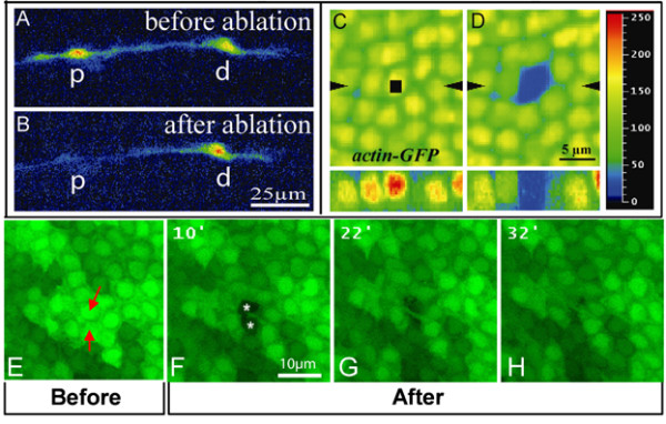
High resolution images of confocal assisted UV laser selective destruction. (a,b) Confocal images of repo::GFP expressing cells prior to (a) and after (b) selective destruction. (b) The proximal glial cell (p) has been targeted for selective destruction; the distal cell (d) was not targeted. (c,d) actin-GFP labeling in the fly wing epithelium prior to (c) and just after (d) UV irradiation. The region of interest defined by the 'Point bleach' function is indicated by the black square in (c). (d) Notice that, upon irradiation, GFP labeling is specifically absent in the targeted nucleus. Images to the bottom of (c,d) correspond to Z optical sections taken along the axis indicated by the black arrowheads. Color coding is used to quantify GFP labeling: blue (0) corresponds to background, red (250) to high levels. (e-h) actin-GFP labeling in the fly wing epithelium prior to (e) and 10 (f), 22 (g) or 32 minutes (h) after UV irradiation. The two targeted cells (indicated by red arrows in (e)) are absent 10 minutes after UV-mediated destruction (indicated by white asterisks in (f)). The space previously occupied by the targeted cells is subsequently occupied by the neighboring cells (g,h). See also Additional file 5. Scale bars: (a,b) 25 μm; (c,d) 5 μm; (e-h) 10 μm.
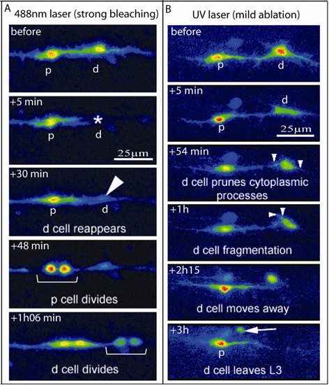
Effects of UV irradiation versus bleaching. (a,b) Color coding of confocal images of repo::GFP expressing cells in strong bleaching (a) or mild UV irradiation (b) experiments. (a) The distal glial cell (d) has been targeted using the 488 nm ray. GFP completely disappears from the cell within 5 minutes after bleaching (asterisk) and subsequently resumes (arrowhead). Notice that both targeted (d) and non-targeted (p) cells divide and that they do it almost simultaneously. See also Additional file 3. (b) The distal cell (d) has been targeted for mild UV irradiation; 54 minutes to 1 hour afterwards, the d cell prunes its cytoplasmic processes and undergoes fragmentation (small arrowheads). Finally, between 2.25 and 3 hours after UV irradiation, the d cell loses contact with its neighbors, acquires a round shape and moves away (arrow). See also Additional file 4. Scale bars: 25 μm.
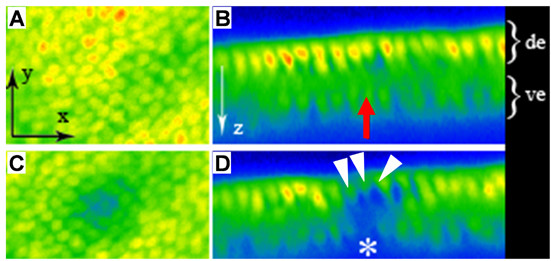
UV cell destruction of ventral wing epithelial cells does not affect dorsal cells. (a-d) GFP labeling prior to (a,b) and after (c,d) UV cell destruction in fly wing epithelium. Dorsal views are shown in (a,c). The wing epithelium presents two cell layers as shown on transversal sections in (b,d). Dorsal epithelium (de) is to the top, ventral epithelium (ve) to the bottom. In each panel, acquisition is made from dorsal to ventral. The same color coding as described in Figure 1 is used to quantify GFP labeling. Before (b) and after (d) irradiation, the ventrally located targeted cell is indicated by a red arrow (b) and a white asterisk (d), respectively. Note that the dorsal GFP-positive cells (white arrowheads in (d)) located just above the ventral targeted cell are still present upon UV irradiation, even though they display a reduced fluorescence signal. See also Additional file 6.
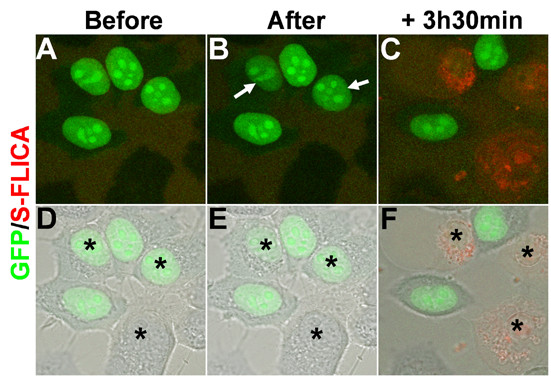
Confocal assisted cell destruction of HeLa cells. (a-f) Confocal images of HeLa cells transfected with pCIG nuclear GFP-expressing vector prior to (a,d), and just after (b,e) cell destruction, and after 3.5 hours of time-lapse analysis (c,f). Before UV irradiation, the S-FLICA apoptosis marker was added to the medium, becoming red in the presence of active caspases. (d-f) Merged images of GFP (green)/S-FLICA (red) colabeling with Nomarski images. Black asterisks indicate the two GFP-positive and the GFP-negative cells that have been targeted for ablation. Note that after 5 seconds of UV irradiation within the nucleus, weak GFP bleaching occurs in targeted GFP-positive cells (white arrows in (b)). After 3.5 hours, all the targeted cells (but not non-targeted cells) express active caspases, as indicated by red labeling. See also Additional file 7.
References
-
- Sweeney TS, Hidalgo A, Belle JSd, Keshishian H. In: Functional ablation. Sullivan B, Ashburner M, Hawley S, editor. Drosophila Protocols Cold Spring Harbor Laboratory Press; 2000. pp. 449–477. - PubMed
Publication types
MeSH terms
Substances
LinkOut - more resources
Full Text Sources
Other Literature Sources
Molecular Biology Databases
