Tetralogy of Fallot with rheumatic mitral stenosis: a case report
- PMID: 18442391
- PMCID: PMC2390577
- DOI: 10.1186/1752-1947-2-127
Tetralogy of Fallot with rheumatic mitral stenosis: a case report
Abstract
Introduction: Rheumatic and congenital heart diseases account for the majority of hospital admissions for cardiac patients in India. Tetralogy of Fallot is the most common congenital heart disease with survival to adulthood. Infective endocarditis accounts for 4% of admissions to a specialized unit for adult patients with a congenital heart lesion. This report is unique in that a severe stenotic lesion of the mitral valve, probably of rheumatic aetiology, was noted in an adult male with Tetralogy of Fallot.
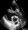
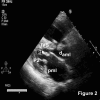
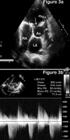
Case presentation: An unusual association of rheumatic mitral stenosis in an adult Indian male patient aged 35 years with Tetralogy of Fallot and subacute bacterial endocarditis of the aortic valve is presented.
Conclusion: In this case report the diagnostic implications, hemodynamic and therapeutic consequences of mitral stenosis in Tetralogy of Fallot are discussed. In addition, the morbidity and mortality of infective endocarditis in adult patients with congenital heart disease are summarized. The risk of a coincident rheumatic process in patients with congenital heart disease is highlighted and the need for careful attention to this possibility during primary and follow-up evaluation of such patients emphasized.
Figures


References
-
- Savitri S. Rheumatic heart disease: is it declining in India? Indian Heart J. 2007;59:9–10. - PubMed
-
- Bokhandi SS, Tullu MS, Shaharao VB, Bavdekar SB, Kamat JR. Congenital heart disease with rheumatic fever and rheumatic heart disease: a coincidence or an association? J Postgrad Med. 2002;48:238–238. - PubMed
-
- Mohan JC, Arora R, Khalilullah M. Double outlet right ventricle with calcified rheumatic mitral stenosis. Indian Heart J. 1991;43:397–399. - PubMed
-
- Kirklin JW, Baratt Boyes BG. Ventricular septal defect with pulmonary stenosis or atresia. In: Kochoukos NT, Blackstone EH, Hanley FL, Doty DB, Karp RB, editor. Cardiac Surgery: Morphology, Diagnostic Criteria, Natural History, Techniques, Results, and Indications. 3. Philadelphia: Churchill Livingstone; 2003. pp. 946–1073.
-
- Perloff JK. Congenital obstruction to left atrial flow: mitral stenosis, cor triatriatum, pulmonary vein stenosis. In: Perloff JK, editor. The Clinical Recognition of Congenital Heart Disease. 5. Philadelphia: Saunders; 2003. pp. 144–156.
LinkOut - more resources
Full Text Sources

