. 2008 Apr 28:9:42.
doi: 10.1186/1471-2202-9-42.

Isolation of neuronal chromatin from brain tissue

Affiliations

Isolation of neuronal chromatin from brain tissue

Yan Jiang et al. BMC Neurosci. .

Abstract

Background: DNA-protein interactions in mature brain are increasingly recognized as key regulators for behavioral plasticity and neuronal dysfunction in chronic neuropsychiatric disease. However, chromatin assays typically lack single cell resolution, and therefore little is known about chromatin regulation of differentiated neuronal nuclei that reside in brain parenchyma intermingled with various types of non-neuronal cells.

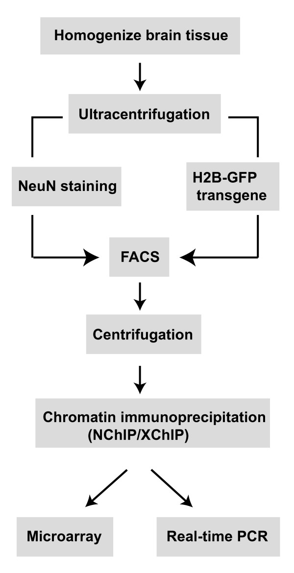
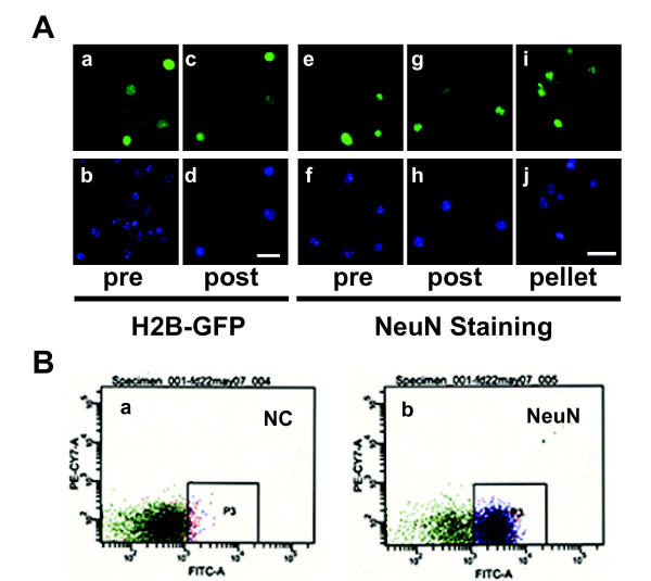
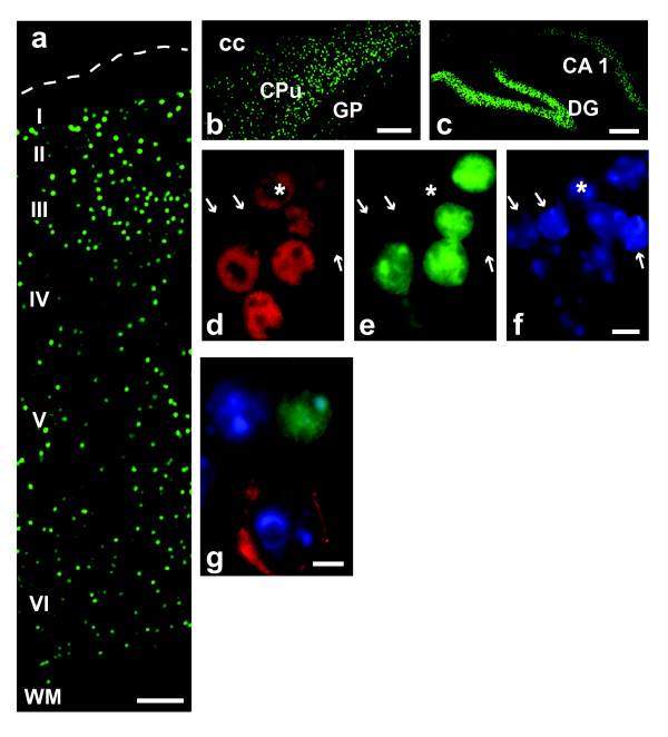
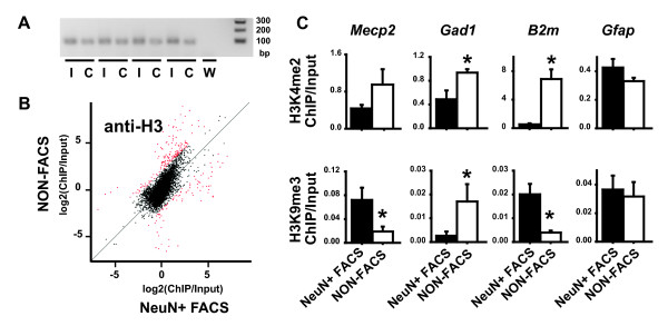
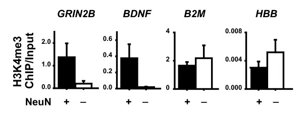
Results: Here, we describe a protocol to selectively tag neuronal nuclei from adult brain - either by (anti-NeuN) immunolabeling or transgene-derived histone H2B-GFP fusion protein - for subsequent fluorescence-activated sorting and chromatin immunoprecipitation (ChIP). To illustrate an example, we compared histone H3 lysine 4 and 9 methylation marks at select gene promoters in neuronal, non-neuronal and unsorted chromatin from mouse forebrain and human cerebral cortex, and provide evidence for neuron-specific histone methylation signatures.

Conclusion: With the modifications detailed in this protocol, the method can be used to collect nuclei from specific subtypes of neurons from any brain region for subsequent ChIP with native/un-fixed or crosslinked chromatin preparations. Starting with the harvest of brain tissue, ChIP-ready neuronal nuclei can be obtained within one day.


Figures

Figure 1
Figure 1
Outline of the procedure to isolate neuronal nuclei from brain for chromatin immunoprecipitation.
Figure 2
Figure 2
Neuronal nuclei isolated from adult brain via FACS. (A) Digitized images of nuclei extracted from forebrain of adult (a-d) CAMIIK-H2B-GFP transgenic mice, and (e-j) wild type mice labeled with NeuN immunoreactivity, as indicated pre and post (FACS sorting), and after pelleting. Green channel for (a, c) GFP or (e, g, i) NeuN; blue channel for DAPI. Notice that post-FACS samples are comprised entirely of neuronal nuclei. Bar = 20 μm. (B) Representative FACS scatter plots from (a) negative control (NC) processed without NeuN antibody, and (b) sample processed with NeuN antibody (NeuN).
Figure 3
Figure 3
Neurons expressing H2B-GFP transgene. Digitized images from 8 week old CAMIIK-H2B-GFP mice. (a) Cerebral cortex. Notice GFP (green) nuclei across layers II-VI, but not in layer I or white matter (WM). (b) caudate-putamen and (c) hippocampus. (d-f) Nuclei extracted from forebrain and processed for NeuN (red), and nucleophilic dye, DAPI (blue). Notice that GFP (green) expression is limited to 5 of the 6 NeuN immunoreactive nuclei. Arrows label GFP-, NeuN- nuclei, * labels GFP-, NeuN+ nucleus. (g) Section from cerebral cortex labeled with anti-GAD67 antibody (red). Notice absence of GFP signal in GABA neuron. Bar is (a, c) 100, (b) 300, (d-g) 5 μm.
Figure 4
Figure 4
Neuron-specific chromatin signatures in mouse brain. (A) Representative gel picture for 25 cycle PCR product (ChIP-GLAS) from 4 independent replicates of Input I and ChIP C, and water control W. (B) Scatter plot showing correlation between Chip-to-input ratios signal from NeuN+ sorted nuclei (NeuN+ FACS) (x-axis) and unsorted nuclei (NON-FACS) (y-axis), using modification-independent antibody against C-terminus of histone H3 anti-H3. (C) Bar graphs showing ChIP-to-input ratio (y-axis) for (top row) dimethylated H3-lysine 4 (H3K4me2) and (bottom row) trimethylated H3-lysine 9 (H3K9me3) as determined for Mecp2, Gad1, B2m, and Gfap gene promoters in chromatin of NeuN+ FACS and NON-FACS nuclei from mouse forebrain. Data shown as mean ± S.E.M., N = 6/sorting; *p < 0.05, ANOVA NeuN+ FACS v.s. NON-FACS.
Figure 5
Figure 5
Neuron-specific chromatin signatures in human prefrontal cortex. Bar graphs showing ChIP-to-input ratio (y-axis) for trimethylated H3-lysine 4 (H3K4me3) at GRIN2B, BDNF and B2M promoters, and HBB (β-globin) locus control region in chromatin of NeuN positive (NeuN+) and negative (NeuN-) FACS sorted nuclei from human prefrontal cortex. Data shown as mean ± S.E.M., N = 3 specimens/sorting. The NeuN+ fraction showed, in comparison to NeuN- fraction of the same subject, increased levels of H3K4me3 at GRIN2B and BDNF in 3/3 cases. Notice further the extremely low ChIP-to-input ratio for HBB.

Similar articles

Cited by

References

    1. Tsankova N, Renthal W, Kumar A, Nestler EJ. Epigenetic regulation in psychiatric disorders. Nat Rev Neurosci. 2007;8:355–367. doi: 10.1038/nrn2132. - DOI - PubMed
    1. Francke U. Mechanisms of disease: neurogenetics of MeCP2 deficiency. Nat Clin Pract Neurol. 2006;2:212–221. doi: 10.1038/ncpneuro0148. - DOI - PubMed
    1. Weaver IC, Cervoni N, Champagne FA, D'Alessio AC, Sharma S, Seckl JR, Dymov S, Szyf M, Meaney MJ. Epigenetic programming by maternal behavior. Nat Neurosci. 2004;7:847–854. doi: 10.1038/nn1276. - DOI - PubMed
    1. Lubin FD, Sweatt JD. The IkappaB kinase regulates chromatin structure during reconsolidation of conditioned fear memories. Neuron. 2007;55:942–957. doi: 10.1016/j.neuron.2007.07.039. - DOI - PMC - PubMed
    1. Nan X, Hou J, Maclean A, Nasir J, Lafuente MJ, Shu X, Kriaucionis S, Bird A. Interaction between chromatin proteins MECP2 and ATRX is disrupted by mutations that cause inherited mental retardation. Proc Natl Acad Sci U S A. 2007;104:2709–2714. doi: 10.1073/pnas.0608056104. - DOI - PMC - PubMed

Publication types

MeSH terms

LinkOut - more resources