

Spatial and temporal distribution patterns of Na-K-2Cl cotransporter in adult and developing mouse retinas
- PMID: 18442435
- PMCID: PMC5531596
- DOI: 10.1017/S0952523808080164
Spatial and temporal distribution patterns of Na-K-2Cl cotransporter in adult and developing mouse retinas
Abstract
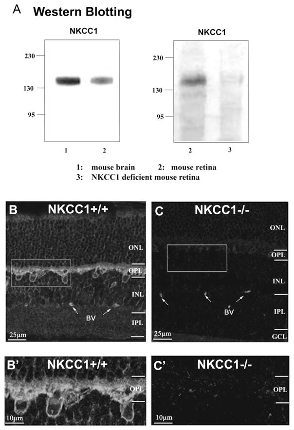
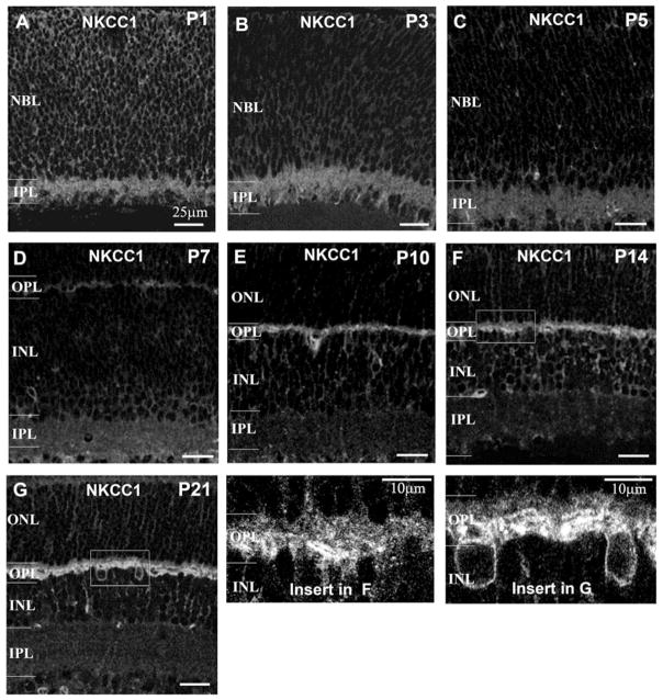
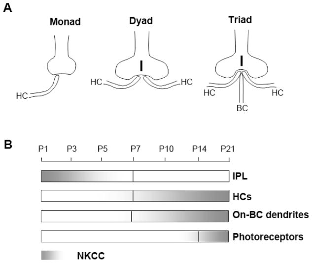
The Na-K-2Cl cotransporter (NKCC) is a Cl(-) uptake transporter that is responsible for maintaining a Cl(-) equilibrium potential positive to the resting potential in neurons. If NKCC is active, GABA and glycine can depolarize neurons. In view of the abundance of GABAergic and glycinergic synapses in retina, we undertook a series of studies using immunocytochemical techniques to determine the distribution of NKCC in retinas of both developing and adult mice. We found NKCC antibody (T4) labeling present in retinas from wild-type mice, but not in NKCC1-deficient mice, suggesting that the NKCC1 subtype is a major Cl(-) uptake transporter in mouse retina. Strong labeling of NKCC1 was present in horizontal cells and rod-bipolar dendrites in adult mice. Interestingly, we also found that a diffuse labeling pattern was present in photoreceptor terminals. However, NKCC1 was barely detectable in the inner retina of adult mice. Using an antibody against K-Cl cotransporter 2 (KCC2), we found that KCC2, a transporter that extrudes Cl(-), was primarily expressed in the inner retina. The expression of NKCC1 in developing mouse retinas was studied from postnatal day (P) 1 to P21, NKCC1 labeling first appeared in the dendrites of horizontal and rod-bipolar cells as early as P7, followed by photoreceptor terminals between P10-P14; with expression gradually increasing concomitantly with the growth of synaptic terminals and dendrites throughout retinal development. In the inner retina, NKCC1 labeling was initially observed in the inner plexiform layer at P1, but labeling diminished after P5. The developmental increase in NKCC expression only occurred in the outer retina. Our results suggest that the distal synapses and synaptogenesis in mouse retinas undergo a unique process with a high intracellular Cl(-) presence due to NKCC1 expression.
Figures


Similar articles
-
Regulation of KCC2 and NKCC during development: membrane insertion and differences between cell types.J Comp Neurol. 2006 Nov 1;499(1):132-43. doi: 10.1002/cne.21100. J Comp Neurol. 2006. PMID: 16958091
-
Localization and developmental expression patterns of the neuronal K-Cl cotransporter (KCC2) in the rat retina.J Neurosci. 2000 Feb 15;20(4):1414-23. doi: 10.1523/JNEUROSCI.20-04-01414.2000. J Neurosci. 2000. PMID: 10662832 Free PMC article.
-
Postnatal maturation of Na+, K+, 2Cl- cotransporter expression and inhibitory synaptogenesis in the rat hippocampus: an immunocytochemical analysis.Eur J Neurosci. 2002 Jan;15(2):233-45. doi: 10.1046/j.0953-816x.2001.01854.x. Eur J Neurosci. 2002. PMID: 11849291
-
The role of Na-K-Cl co-transporter in cerebral ischemia.Neurol Res. 2005 Apr;27(3):280-6. doi: 10.1179/016164105X25243. Neurol Res. 2005. PMID: 15845211 Review.
-
The bumetanide-sensitive Na-K-2Cl cotransporter NKCC1 as a potential target of a novel mechanism-based treatment strategy for neonatal seizures.Neurosurg Focus. 2008 Sep;25(3):E22. doi: 10.3171/FOC/2008/25/9/E22. Neurosurg Focus. 2008. PMID: 18759624 Review.
Cited by
-
Calcium channels in rat horizontal cells regulate feedback inhibition of photoreceptors through an unconventional GABA- and pH-sensitive mechanism.J Physiol. 2013 Jul 1;591(13):3309-24. doi: 10.1113/jphysiol.2012.248179. Epub 2013 Apr 22. J Physiol. 2013. PMID: 23613534 Free PMC article.
-
Mechanisms of chloride uptake in frog olfactory receptor neurons.J Comp Physiol A Neuroethol Sens Neural Behav Physiol. 2011 Apr;197(4):339-49. doi: 10.1007/s00359-010-0618-1. Epub 2011 Jan 21. J Comp Physiol A Neuroethol Sens Neural Behav Physiol. 2011. PMID: 21253748
-
Role of the neuronal K-Cl co-transporter KCC2 in inhibitory and excitatory neurotransmission.Front Cell Neurosci. 2012 Feb 21;6:5. doi: 10.3389/fncel.2012.00005. eCollection 2012. Front Cell Neurosci. 2012. PMID: 22363264 Free PMC article.
-
GABAergic signalling in a neurogenic niche of the turtle spinal cord.J Physiol. 2011 Dec 1;589(Pt 23):5633-47. doi: 10.1113/jphysiol.2011.214312. Epub 2011 Sep 12. J Physiol. 2011. PMID: 21911613 Free PMC article.
-
Cellular Physiology and Pathophysiology of EAAT Anion Channels.Front Cell Neurosci. 2022 Jan 6;15:815279. doi: 10.3389/fncel.2021.815279. eCollection 2021. Front Cell Neurosci. 2022. PMID: 35087380 Free PMC article. Review.
References
-
- Balkema GW, Rizkalla R. Ultrastructural localization of a synaptic ribbon protein recognized by antibody B16. Journal of Neurocytology. 1996;25:565–571. - PubMed
-
- Beltran WA, Rohrer H, Aguirre GD. Immunolocalization of ciliary neurotrophic factor receptor alpha (CNTFRalpha) in mammalian photoreceptor cells. Molecular Vision. 2005;11:232–244. - PubMed
-
- Ben-Ari Y. Excitatory actions of GABA during development: The nature of the nurture. Nature Reviews Neurosciences. 2002;3:728–739. - PubMed
-
- Blanco R, Vaquero CF, de la Villa P. The effects of GABA and glycine on horizontal cells of the rabbit retina. Vision Research. 1996;36:3987–3995. - PubMed
-
- Blanks JC, Adinolfi AM, Lolley RN. Synaptogenesis in the photoreceptor terminal of the mouse retina. Journal of Comparative Neurology. 1974;156:81–93. - PubMed
Publication types
MeSH terms
Substances
Grants and funding
LinkOut - more resources
Full Text Sources
Other Literature Sources
Medical
Research Materials
Miscellaneous
