

Quantitative expression patterns of peroxisome proliferator-activated receptor-beta/delta (PPARbeta/delta) protein in mice
- PMID: 18442472
- PMCID: PMC2586836
- DOI: 10.1016/j.bbrc.2008.04.086
Quantitative expression patterns of peroxisome proliferator-activated receptor-beta/delta (PPARbeta/delta) protein in mice
Abstract
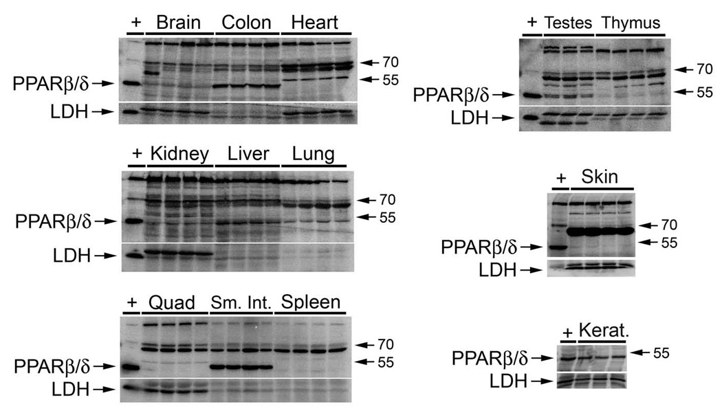
The expression patterns of PPARbeta/delta have been described, but the majority of these data are based on mRNA data. To date, there are no reports that have quantitatively examined the expression of PPARbeta/delta protein in mouse tissues. In the present study, a highly specific PPARbeta/delta antibody was developed, characterized, and used to examine tissue expression patterns of PPARbeta/delta. As compared to commercially available anti-PPARbeta/delta antibodies, one of six polyclonal anti-PPARbeta/delta antibodies developed was significantly more effective for immunoprecipitation of in vitro-translated PPARbeta/delta. This antibody was used for quantitative Western blot analysis using radioactive detection methods. Expression of PPARbeta/delta was highest in colon, small intestine, liver, and keratinocytes as compared to other tissues including heart, spleen, skeletal muscle, lung, brain, and thymus. Interestingly, PPARbeta/delta expression was localized in the nucleus and RXRalpha can be co-immunoprecipitated with nuclear PPARbeta/delta. Results from these studies demonstrate that PPARbeta/delta expression is highest in intestinal epithelium, liver, and keratinocytes, consistent with significant biological roles in these tissues.
Figures


Similar articles
-
Functional characterization of peroxisome proliferator-activated receptor-β/δ expression in colon cancer.Mol Carcinog. 2011 Nov;50(11):884-900. doi: 10.1002/mc.20757. Epub 2011 Mar 11. Mol Carcinog. 2011. PMID: 21400612 Free PMC article.
-
The coactivator PGC-1α regulates skeletal muscle oxidative metabolism independently of the nuclear receptor PPARβ/δ in sedentary mice fed a regular chow diet.Diabetologia. 2014 Nov;57(11):2405-12. doi: 10.1007/s00125-014-3352-3. Epub 2014 Aug 13. Diabetologia. 2014. PMID: 25116175 Free PMC article.
-
Regulation of Cytochrome P450 2B10 (CYP2B10) Expression in Liver by Peroxisome Proliferator-activated Receptor-β/δ Modulation of SP1 Promoter Occupancy.J Biol Chem. 2016 Nov 25;291(48):25255-25263. doi: 10.1074/jbc.M116.755447. Epub 2016 Oct 20. J Biol Chem. 2016. PMID: 27765815 Free PMC article.
-
Dissecting the role of peroxisome proliferator-activated receptor-β/δ (PPARβ/δ) in colon, breast, and lung carcinogenesis.Cancer Metastasis Rev. 2011 Dec;30(3-4):619-40. doi: 10.1007/s10555-011-9320-1. Cancer Metastasis Rev. 2011. PMID: 22037942 Free PMC article. Review.
-
Peroxisome proliferator-activated receptor beta/delta as a therapeutic target for metabolic diseases.Expert Opin Ther Targets. 2005 Aug;9(4):861-73. doi: 10.1517/14728222.9.4.861. Expert Opin Ther Targets. 2005. PMID: 16083348 Review.
Cited by
-
Xenobiotic-sensing nuclear receptors involved in drug metabolism: a structural perspective.Drug Metab Rev. 2013 Feb;45(1):79-100. doi: 10.3109/03602532.2012.740049. Epub 2012 Dec 5. Drug Metab Rev. 2013. PMID: 23210723 Free PMC article. Review.
-
Ligand activation of peroxisome proliferator-activated receptor-beta/delta and inhibition of cyclooxygenase-2 enhances inhibition of skin tumorigenesis.Toxicol Sci. 2010 Jan;113(1):27-36. doi: 10.1093/toxsci/kfp212. Epub 2009 Sep 11. Toxicol Sci. 2010. PMID: 19748995 Free PMC article.
-
Ligand activation of peroxisome proliferator-activated receptor beta/delta (PPAR beta/delta) inhibits chemically induced skin tumorigenesis.Carcinogenesis. 2008 Dec;29(12):2406-14. doi: 10.1093/carcin/bgn219. Epub 2008 Sep 17. Carcinogenesis. 2008. PMID: 18799709 Free PMC article.
-
Unveiling behavioral and molecular neuroadaptations related to the antidepressant action of cannabidiol in the unpredictable chronic mild stress model.Front Pharmacol. 2023 Apr 18;14:1171646. doi: 10.3389/fphar.2023.1171646. eCollection 2023. Front Pharmacol. 2023. PMID: 37144214 Free PMC article.
-
PFAS and Potential Adverse Effects on Bone and Adipose Tissue Through Interactions With PPARγ.Endocrinology. 2021 Dec 1;162(12):bqab194. doi: 10.1210/endocr/bqab194. Endocrinology. 2021. PMID: 34480479 Free PMC article. Review.
References
-
- Grimaldi PA. Regulatory functions of PPARbeta in metabolism: implications for the treatment of metabolic syndrome. Biochim Biophys Acta. 2007;1771:983–990. - PubMed
-
- Burdick AD, Kim DJ, Peraza MA, Gonzalez FJ, Peters JM. The role of peroxisome proliferator-activated receptor-beta/delta in epithelial cell growth and differentiation. Cell Signal. 2006;18:9–20. - PubMed
-
- Braissant O, Foufelle F, Scotto C, Dauca M, Wahli W. Differential expression of peroxisome proliferator-activated receptors (PPARs): tissue distribution of PPAR-alpha, -beta, and -gamma in the adult rat. Endocrinology. 1996;137:354–366. - PubMed
Publication types
MeSH terms
Substances
Grants and funding
LinkOut - more resources
Full Text Sources
Other Literature Sources
Molecular Biology Databases
Research Materials
