. 2008 Jul 11;283(28):19176-83.
doi: 10.1074/jbc.M802299200. Epub 2008 Apr 28.

Regulation of intra-S phase checkpoint by ionizing radiation (IR)-dependent and IR-independent phosphorylation of SMC3

Affiliations

Regulation of intra-S phase checkpoint by ionizing radiation (IR)-dependent and IR-independent phosphorylation of SMC3

Hao Luo et al. J Biol Chem. .

Abstract

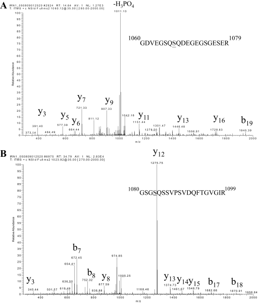
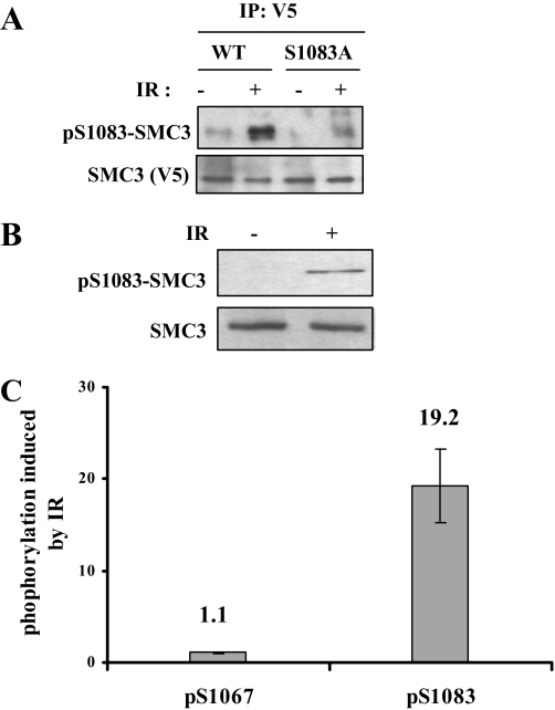
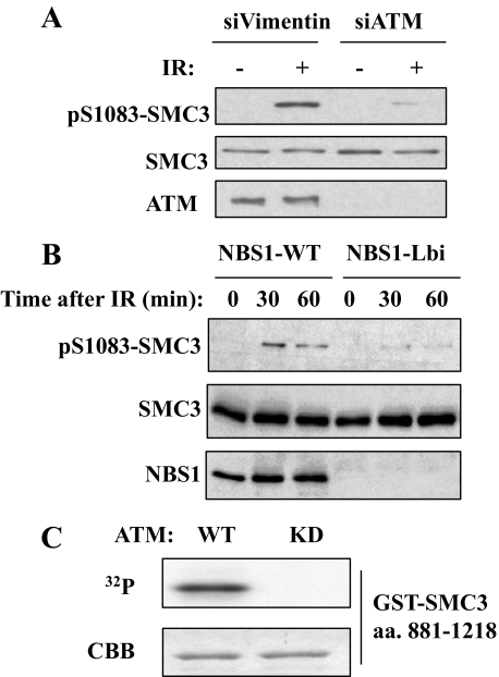
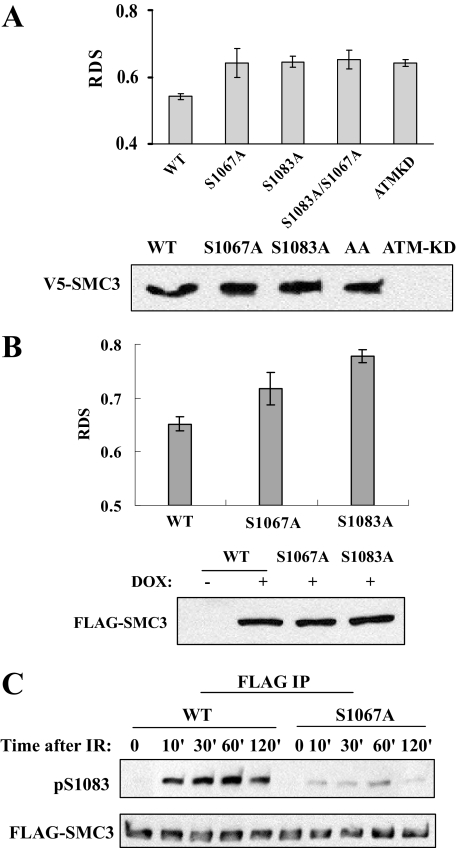
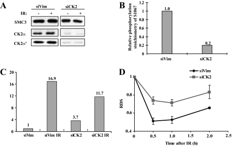
Structure maintenance of chromosome 1 (SMC1) is phosphorylated by ataxia telangiectasia-mutated (ATM) in response to ionizing radiation (IR) to activate intra-S phase checkpoint. A role of CK2 in DNA damage response has been implicated in many previous works, but the molecular mechanism for its activation is not clear. In the present work, we report that SMC3 is phosphorylated at Ser-1067 and Ser-1083 in vivo. Ser-1083 phosphorylation is IR-inducible, depends on ATM and Nijmegen breakage syndrome 1 (NBS1), and is required for intra-S phase checkpoint. Interestingly, Ser-1067 phosphorylation is constitutive and is not induced by IR but also affects intra-S phase checkpoint. Phosphorylation of Ser-1083 is weakened in cells expressing S1067A mutant, suggesting interplay between Ser-1067 and Ser-1083 phosphorylation in DNA damage response. Consistently, small interfering RNA knockdown of CK2 leads to attenuated phosphorylation of Ser-1067 as well as intra-S phase checkpoint defect. Our data provide evidence that phosphorylation of a core cohesin subunit SMC3 by ATM plays an important role in DNA damage response and suggest that a constitutive phosphorylation by CK2 may affect intra-S phase checkpoint by modulating SMC3 phosphorylation by ATM.


Figures

FIGURE 1.
FIGURE 1.
SMC3 is phosphorylated at Ser-1067 and Ser-1083. MS/MS spectra that identify Ser-1067 (A) and Ser-1083 (B) as phosphorylated. SMC3 was immunoprecipitated from nuclear extracts made from HeLa cells and further isolated on SDS-PAGE. The SMC3 band was in-gel digested with trypsin and analyzed with capillary liquid chromatography-MS/MS.
FIGURE 2.
FIGURE 2.
SMC3 is phosphorylated at Ser-1067 and Ser-1083 in cycling and IR-treated cells. A, characterization of pS1083-SMC3 phospho-specific antibody. V5-SMC3-WT and V5-SMC3-S1083A were transfected in 293T cells, and cells were irradiated with 10 Gy IR. SMC3 was immunoprecipitated (IP) with a V5 antibody and Western blotted with pS1083-SMC3 antibody. B, phosphorylation of the endogenous SMC3 at Ser-1083. HeLa cells were irradiated with 10 Gy of IR and allowed to recover for 1 h. Whole cell lysates were Western blotted with antibodies against pS1083-SMC3 and SMC3. C, quantitative mass spectrometric measurement of Ser-1067 and Ser-1083 phosphorylation induction in response to IR. HeLa cells were labeled with light or heavy isotope and treated with or without IR, respectively. Equal amounts of two isotope-labeled cells were mixed for the subsequent processing. Phosphorylation of Ser-1067 and Ser-1083 in light labeled SMC3 (IR-treated) are normalized to those in heavy labeled SMC3 (untreated) to calculate phosphorylation induced by IR.
FIGURE 3.
FIGURE 3.
The ATM-NBS1 pathway regulates Ser-1083 phosphorylation of SMC3 in response to IR. A, Western blotting of SMC3 phosphorylation of Ser-1083 in siVimentin- and siATM-transfected in HeLa cells that were irradiated with 10 Gy IR and recovered for 1 h. B, requirement of NBS1 and its phosphorylation for IR-induced phosphorylation of SMC3 at Ser-1083. NBS cells complemented with NBC1 (NBS1-WT) or an empty vector (NBS1-Lbi) were treated with 10 Gy of IR and harvested at the indicated times and Western blotted for SMC3 Ser-1083 phosphorylation. C, in vitro phosphorylation of SMC3 by ATM. FLAG-ATM overexpressed in 293T cells was immunoprecipitated by anti-FLAG antibody and incubated with a GST-SMC3 fragment (amino acids 881–1218) and γ-32P-labeled ATP. In vitro phosphorylation was detected by autoradiography. The amount of SMC3 fragment on SDS-PAGE gel was shown by Coomassie Brilliant Blue (CBB) staining. KD, kinase-dead.
FIGURE 4.
FIGURE 4.
Phosphorylation of both Ser-1067 and Ser-1083 is required for intra-S phase checkpoint activation. A, RDS measurements of cells transiently transfected with WT, mutant SMC3, or kinase-dead ATM (ATMKD) genes. Plasmids encoding genes of interest were transfected into 293T cells with Lipofectamine™, and cells were labeled with 14C 48 h after transfection. Cells were treated with 10 Gy IR and allowed to recover for 1 h. RDS was measured as described under “Materials and Methods.” Western blotting by the V5 antibody indicates the expression levels of exogenous WT and mutant V5-SMC3. B, RDS measurements of FLAG-SMC3 stable cell lines. WT or mutant FLAG-SMC3 cell lines were cultured in Dulbecco's modified Eagle's medium with doxycycline (DOX) for 24 h before 14C labeling. Cells were treated with 10 Gy IR and allowed to recover for 1 h. RDS was measured as described under “Materials and Methods.” Western blotting by the FLAG antibody was used to determine the expression levels of exogenous WT and mutant FLAG-SMC3 after doxycycline addition for 24 h. AA, S1083A/S1067A. C, SMC3 Ser-1083 phosphorylation in a S1067A stable cell line. FLAG-SMC3-WT and -S1067A-expressing 293T cell lines were treated with 5 Gy of IR and recovered for the indicated times. WT and mutant FLAG-SMC3 were immunoprecipitated (IP) from cells lysates with anti-FLAG M2 antibody and Western blotted with phospho-Ser-1083 or FLAG antibody.
FIGURE 5.
FIGURE 5.
CK2 is required for phosphorylation of Ser-1067 and regulates Ser-1083 phosphorylation in response to IR and intra-S phase checkpoint. A, CK2 is knocked down by co-transfection of siRNA against both α and α′ subunits. B, quantitative mass spectrometric measurements of Ser-1067 phosphorylation. SMC3 was immunoprecipitated from siCK2 (both CK2α and CK2α′ was knocked down) and siVim (as a control)-transfected cells. The percentage of Ser-1067 phosphorylation was normalized to that in the siVim cell. C, quantitative mass spectrometric measurements of Ser-1083 phosphorylation. SMC3 was purified from siCK2 and siVim cells with or without IR treatment. Phosphorylation of Ser-1083 in indicated conditions was measured and normalized to the level in siVim-transfected cells without IR. D, RDS measurements in siCK2 cells and siVim cells. Sixty hours after siRNA transfection, HeLa cells were labeled with 14C for 36 h and then treated with 10 Gy IR. RDS was measured at the indicated times.

References

    1. Zhou, B. B., and Elledge, S. J. (2000) Nature 408 433–439 - PubMed
    1. Kastan, M. B., and Bartek, J. (2004) Nature 432 316–323 - PubMed
    1. Litchfield, D. W. (2003) Biochem. J. 369 1–15 - PMC - PubMed
    1. Homma, M. K., and Homma, Y. (2005) Mol. Cell Biochem. 274 47–52 - PubMed
    1. Harvey, E. J., Li, N., and Ramji, D. P. (2007) Arterioscler. Thromb. Vasc. Biol. 27 806–812 - PubMed

Publication types

MeSH terms