. 2008 Jul;76(7):2978-90.
doi: 10.1128/IAI.00075-08. Epub 2008 Apr 28.

Hemolysin of uropathogenic Escherichia coli evokes extensive shedding of the uroepithelium and hemorrhage in bladder tissue within the first 24 hours after intraurethral inoculation of mice

Affiliations

Hemolysin of uropathogenic Escherichia coli evokes extensive shedding of the uroepithelium and hemorrhage in bladder tissue within the first 24 hours after intraurethral inoculation of mice

Yarery C Smith et al. Infect Immun. 2008 Jul.

Abstract

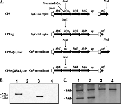
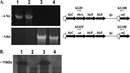
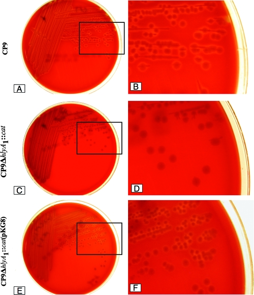
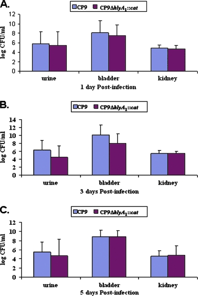
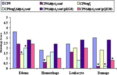
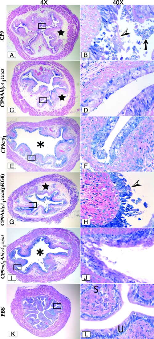
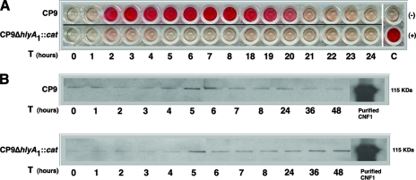
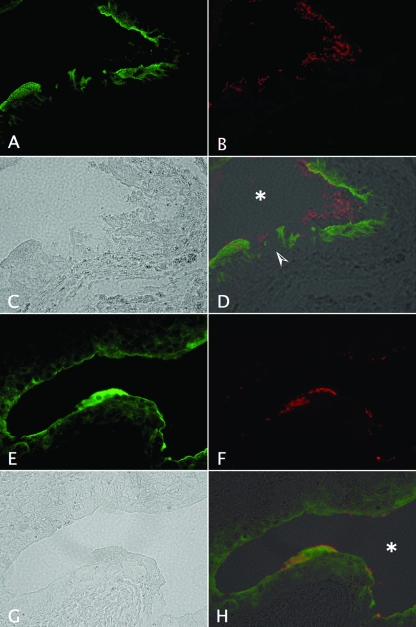
Many uropathogenic Escherichia coli (UPEC) strains produce both hemolysin (Hly) and cytotoxic necrotizing factor type 1 (CNF1), and the loci for these toxins are often linked. The conclusion that Hly and CNF1 contribute to urovirulence is supported by the results of epidemiological studies associating the severity of urinary tract infections (UTIs) with toxin production by UPEC isolates. Additionally, we previously reported that mouse bladders and rat prostates infected with UPEC strain CP9 exhibit a more profound inflammatory response than the organs from animals challenged with CP9cnf(1) and that CNF1 decreases the antimicrobial activities of polymorphonuclear leukocytes. More recently, we created an Hly mutant, CP9Delta hlyA(1)::cat, and showed that it was less hemolytic and destructive for cultured bladder cells than CP9 was. Here we evaluated the relative effects of mutations in hlyA(1) or cnf(1) alone or together on the pathogenicity of CP9 in a mouse model of ascending UTI. To do this, we constructed an hlyA(1)-complemented clone of CP9Delta hlyA(1)::cat and an hlyA(1) cnf(1) CP9 double mutant. We found that Hly had no influence on bacterial colonization of the bladder or kidneys in single or mixed infections with the wild type and CP9Delta hlyA(1)::cat but that it did provoke sloughing of the uroepithelium and bladder hemorrhage within the first 24 h after challenge. Finally, we confirmed that CNF1 expression induces bladder inflammation and, in particular, as shown in this study, submucosal edema. From these data, we speculate that Hly and CNF1 may be largely responsible for the signs and symptoms of cystitis in humans infected with toxigenic UPEC.


Figures

FIG. 1.
FIG. 1.
Diagram of the CP9 hly operon upstream of cnf1 and Southern blot analyses. (A) The mutated or wild-type Hly operon, the intergenic sequence (igs), and the cnf1 gene from the chromosome of strains CP9, CP9cnf1, CP9ΔhlyA1::cat, and CP9cnf1ΔhlyA1::cat are indicated by large filled arrows. Promoter regions (open arrows) are also shown. Sites of NcoI cleavage (indicated by vertical arrows) of chromosomal DNA were predicted from database sequences (J96 hly operon, GenBank accession no. M10133; cnf1, GenBank accession no. X70670; pACYC184 cat gene, GenBank accession no. X06403; J96 igs, GenBank accession no. X70670). (B) Southern blot to detect the NcoI-NcoI cnf1 fragment. Lane 1, CP9; lane 2, CP9cnf1; lane 3, CP9ΔhlyA1::cat; lane 4, CP9cnf1ΔhlyA1::cat. As expected, the fragment of the chromosome that contained the cnf1 gene was discernible in strains CP9 and CP9ΔhlyA1::cat (lanes 1 and 3, respectively). The cnf1-specific band size (∼7.7 kb) was reduced (∼7.0 kb) in the CP9ΔhlyA1::cat strain because hlyA1 was replaced by the smaller cat gene. A cnf1-specific band was absent in the cnf1 mutants (lanes 2 and 4) because the cnf1 probe (KG11F and KG11R) binds to a region of cnf1 that is absent in the mutant strains (due to internal deletion of a 1.2-kb BclI fragment). (C) Southern blot to detect the NcoI-NcoI hlyA fragment(s). Lane 1, CP9; lane 2, CP9cnf1; lane 3, CP9ΔhlyA1::cat; lane 4, CP9cnf1ΔhlyA1::cat. As anticipated, two copies of hlyA were evident in strains CP9 and CP9cnf1 (lanes 1 and 2, respectively), but only one band was apparent in the hlyA1 mutants (lanes 3 and 4) due to deletion of one hlyA gene locus.
FIG. 2.
FIG. 2.
Long-range PCR. (A) Primers KG09 and KG10R (arrows above the hemolysin operon) were used to test for the presence of an hlyA1-containing PCR fragment amplified from hlyA1 to the cnf1 gene on the chromosome of the following strains: CP9 (lane 1), CP9cnf1 (lane 2), CP9ΔhlyA1::cat (lane 3), and CP9cnf1ΔhlyA1::cat (lane 4). PCR products (∼6.7 kb) were present for CP9 and CP9cnf1 but not for CP9ΔhlyA1::cat and CP9cnf1ΔhlyA1::cat, as shown in the top panel of the ethidium bromide-stained agarose gel on the left. Primers KG03F and KG10R were used to verify the replacement of hlyA1 with cat upstream of the cnf1 gene in mutant strains CP9ΔhlyA1::cat and CP9cnf1ΔhlyA1::cat (∼5.0-kb amplified PCR product from cat to cnf1 in the bottom panel of the ethidium bromide-stained agarose gel). (B) Western blot analysis with polyclonal anti-CNF1 serum as a probe was used to verify that wild-type levels of CNF1 (lane 1) were produced by CP9ΔhlyA1::cat (lane 3) and that, as expected, strains CP9cnf1 and CP9cnf1ΔhlyA1::cat did not produce CNF1 (lanes 2 and 4, respectively).
FIG. 3.
FIG. 3.
Blood agar plate assay for hemolytic activity. (A) CP9 plate. (B) Enlargement of the section indicated by a box on the CP9 plate. (C) CP9ΔhlyA1::cat plate. (D) Enlargement of the section indicated by a box on the CP9ΔhlyA1::cat plate. (E) CP9ΔhlyA1::cat(pKG8) plate. (F) Enlargement of the section indicated by a box on the CP9ΔhlyA1::cat(pKG8) plate. Strains were streaked on plates, incubated overnight at 37°C, and photographed digitally with a Nikon Coolpix 995 camera.
FIG. 4.
FIG. 4.
Kinetics of UTI of C3H/HeOuJ mice with CP9 and the CP9ΔhlyA1::cat isogenic mutant. Fifteen female C3H/HeOuJ mice were inoculated via the urethra with 2.5 × 10⁷ CFU of either CP9 or CP9ΔhlyA1::cat. After 1, 3, and 5 days, mice were euthanized and urine samples, as well as bladders and kidneys, were collected, homogenized, and plated on LB agar for bacterial enumeration. (A) Log CFU/ml for 1 day postinfection. (B) Log CFU/ml for 3 days postinfection. (C) Log CFU/ml for 5 days postinfection. Each bar indicates the geometric mean for a sample, and the error bars indicate one standard deviation above the geometric mean.
FIG. 5.
FIG. 5.
Effect of CP9 strains on the histology of murine bladders. Groups of five female C3H/HeOuJ mice were inoculated via the urethra with 2.5 × 10⁷ CFU of CP9, CP9ΔhlyA1::cat, CP9cnf1, CP9cnf1ΔhlyA1::cat, CP9ΔhlyA1::cat(pKG8), or CP9ΔhlyA1::cat(pQE30) and euthanized 1 day later. Bladders were removed, fixed in formalin, and processed for histological examination. Each sample was examined by light microscopy. A score of 0 to 5 was assigned to each bladder for edema, hemorrhage, leukocyte presence, and urothelial damage. Scores for each group of mice were combined, and the averages are indicated by the bars. An asterisk indicates that the tissue response to a strain was statistically significantly different from the response to the strain for which the response was greatest for the specific effect, as determined by ANOVA assessment followed by Tukey's post hoc pairwise comparisons.
FIG. 6.
FIG. 6.
Infection of female C3H/HeOuJ mice with CP9 UPEC strains. Mice were inoculated with CP9 (A and B), CP9ΔhlyA1::cat (C and D), CP9cnf1 (E and F), CP9ΔhlyA1::cat(pKG8) (G and H), and CP9cnf1ΔhlyA1::cat (I and J) for 1 day, and then sections were cut, fixed with formalin, embedded in paraffin, stained with Giemsa stain, and analyzed by light microscopy. Images of a representative sample for each infection are shown at a magnification of ×4 on the left. The rectangles in the images indicate the sections that are shown at a magnification of ×40 on the right. The stars in panels A, C, and G indicate a high degree of edema. The asterisks in panels E and I indicate the luminal space. Note the presence of many inflammatory cells (arrow) in the luminal space in panel B. The arrowheads in panels B and H indicate the intense damage to the urothelium caused by CP9 and CP9ΔhlyA1::cat(pKG8), respectively. In contrast, the minimal damage caused by CP9ΔhlyA1::cat, CP9cnf1, and CP9cnf1ΔhlyA1::cat is shown in panels D, F, and J, respectively. Panels K and L show a murine bladder instilled with only PBS. U and S in panel L indicate the murine urothelium and submucosa, respectively.
FIG. 7.
FIG. 7.
Infection of female C3H/HeOuJ mice with the CP9 UPEC strain after 3 and 5 days. Groups of five mice were inoculated with CP9, and bladders were harvested after 3 days (A and B) and 5 days (C and D) and then processed as described in the legend to Fig. 6. Images of a representative sample for each infection are shown at a magnification of ×4 on the left. The rectangles in the images indicate the sections that are shown at a magnification of ×40 on the right. Panels E and F show a murine bladder instilled with only PBS and harvested after 3 days. U and S in panel F indicate the murine urothelium and submucosa, respectively. The arrows in panels A and C indicate the degree of interstitial edema at 3 and 5 days, respectively.
FIG. 8.
FIG. 8.
Alpha-hemolytic activity and CNF1 expression in CP9 strains. Samples were prepared and assays were carried out as described in Material and Methods. (A) Alpha-hemolytic activity of CP9 or CP9ΔhlyA1::cat supernatants collected hourly from 0 to 48 h was measured by mixing the samples with sheep RBCs. Results for 9 to 17 and 25 to 48 h are not shown. C(−) and C(+) were the negative and positive controls, respectively. (B) Western blot analyses of CP9 and CP9ΔhlyA1::cat bacterial cell lysates probed with polyclonal anti-CNF1 serum.
FIG. 9.
FIG. 9.
Impact of CP9 and the isogenic CP9ΔhlyA1::cat mutant on bladder uroepithelium as revealed by immunostaining of uroplakins. Female C3H/HeOuJ mice were inoculated via the urethra with 2.5 × 10⁷ CFU of CP9 (A to D) or CP9ΔhlyA1::cat (E to H). Mice were euthanized 1 day later. Bladders were removed, fixed in formalin, embedded in paraffin, processed for immunostaining, and analyzed by immunofluorescence microscopy. The sequence shows samples stained with goat anti-uroplakin III antibody (green) (A and E) and with rabbit anti-E. coli antibody (red) (B and F), as well as phase-contrast (C and G) and merged images (D and H). The arrowhead in panel D indicates the severe damage to the uroplakin-lined urothelium caused by CP9, in contrast to the minimal effect on the uroplakin-stained uroepithelium of the CP9ΔhlyA1::cat-infected bladder in panel H. The asterisks in panels D and H indicate the murine bladder luminal space. Magnification, ×40.

References

    1. Aktories, K. 1997. Rho proteins: targets for bacterial toxins. Trends Microbiol. 5282-288. - PubMed
    1. Alonso, P., J. Blanco, M. Blanco, and E. A. Gonzalez. 1987. Frequent production of toxins by Escherichia coli strains isolated from human urinary tract infections: relation with haemagglutination. FEMS Microbiol. Lett. 48391-396.
    1. Andreu, A., A. E. Stapleton, C. Fennell, H. A. Lockman, M. Xercavins, F. Fernandez, and W. E. Stamm. 1997. Urovirulence determinants in Escherichia coli strains causing prostatitis. J. Infect. Dis. 176464-469. - PubMed
    1. Blanco, J., M. P. Alonso, E. A. Gonzalez, M. Blanco, and J. I. Garabal. 1990. Virulence factors of bacteraemic Escherichia coli with particular reference to production of cytotoxic necrotising factor (CNF) by P-fimbriate strains. J. Med. Microbiol. 31175-183. - PubMed
    1. Blanco, J., M. Blanco, M. P. Alonso, J. E. Blanco, E. A. Gonzalez, and J. I. Garabal. 1992. Characteristics of haemolytic Escherichia coli with particular reference to production of cytotoxic necrotizing factor type 1 (CNF1). Res. Microbiol. 143869-878. - PubMed

Publication types

MeSH terms

LinkOut - more resources