. 2008 Jun;10(3):244-53.
doi: 10.1215/15228517-2008-016. Epub 2008 Apr 28.

REIC/Dkk-3 induces cell death in human malignant glioma

Affiliations

REIC/Dkk-3 induces cell death in human malignant glioma

Yoshifumi Mizobuchi et al. Neuro Oncol. 2008 Jun.

Abstract

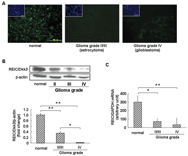
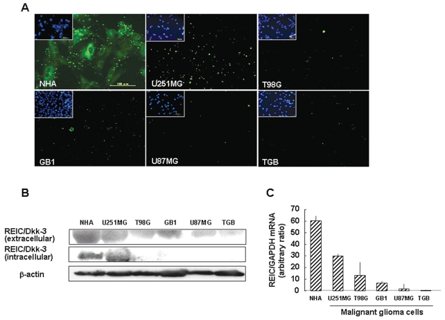
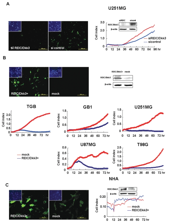
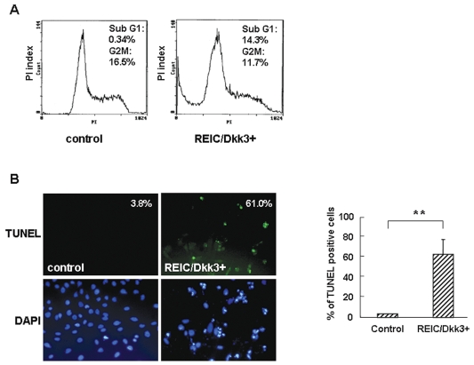
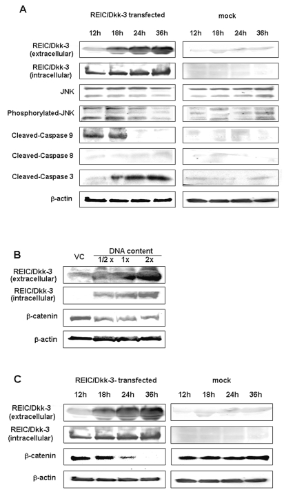
The progression of glioma to more malignant phenotypes results from the stepwise accumulation of genetic alterations and the consequent disruption of the apoptotic pathway and augmentation of survival signaling. REIC/Dkk-3, a member of the human Dickkopf (Dkk) family, plays a role as a suppressor of the growth of several human cancers; however, to date it has not been identified in brain tumors. We compared the gene and protein expression of REIC/Dkk-3 in human malignant glioma and normal brain tissues using quantitative real-time PCR, Western blotting, and immunohistochemistry. We also performed small interfering REIC/Dkk-3 (siREIC/Dkk-3) knockdown and REIC/Dkk-3 overexpression experiments to examine the role of REIC/Dkk-3 in human malignant glioma cells in vitro. In brain tissue from patients with malignant glioma, the gene and protein expression of REIC/Dkk-3 was lower than in normal brain tissue and was related to the malignancy grade. In the primary glioblastoma cell line, REIC/Dkk-3 transfection led to apoptosis owing to the activation of phosphorylated JUN, caspase-9, and caspase-3 and the reduction of beta-catenin; in REIC/Dkk-3 knockdown experiments, cell growth was augmented. Our results suggest that REIC/Dkk-3 regulates the growth and survival of these cells in a caspase-dependent and -independent way via modification of the Wnt signaling pathway. Our work is the first documentation that the gene and protein expression of REIC/Dkk-3 is down-regulated in human malignant glioma. Our demonstration of the mechanisms underlying REIC/Dkk-3-induced cell death indicates that REIC/Dkk-3 plays a pivotal role in the biology of human malignant glioma and suggests that REIC/Dkk-3 is a promising candidate for molecular target therapy.


Figures

Fig. 1
Fig. 1
Protein and mRNA expression of REIC/Dkk-3 in human brain tissue samples. (A) Immunofluorescence staining for REIC/Dkk-3 protein (green) in control temporal lobectomy specimens and astrocytoma and glioblastoma samples. The cells were counterstained for nuclear DNA (blue). The controls stained more strongly and diffusely for REIC/Dkk-3 protein than did glioblastoma, and the degree of staining was correlated with the malignancy grade. (B) The protein level of REIC/Dkk-3 was analyzed in three control lobectomy specimens, three astrocytomas (glioma grades II and III), and three glioblastomas (glioma grade IV) by Western blot analysis, and the protein band density was calculated using Image-J software. Taking the average REIC/Dkk-3 protein level in the three controls as 1.00, the average level was 0.36 in the three astrocytomas (p < 0.05) and 0.02 in the three glioblastomas (p < 0.05), indicating that the higher the WHO glioma grade, the lower the expression of REIC/Dkk-3 protein. (C) Using real-time PCR, we estimated the expression level of REIC/Dkk-3 mRNA in seven control lobectomy specimens, eight astrocytomas, and six glioblastomas. The expression level of REIC/Dkk-3 mRNA relative to glyceraldehyde-3-phosphate dehydrogenase (GAPDH) was significantly lower in glioblastoma than in the controls (p < 0.01). The decrease in REIC/Dkk-3 mRNA expression was inversely correlated with the glioma grade.
Fig. 2
Fig. 2
Protein and mRNA expression of REIC/Dkk-3 in malignant glioma cell lines. (A) Immunofluorescence staining for REIC/Dkk-3 protein (green) in malignant glioma cell lines U251MG, T98G, GB1, U87MG, and TGB and in normal human astrocytes (NHA). NHA cells stained more strongly and diffusely than did the malignant glioma cell lines. U251MG cells stained most strongly; U87MG and TGB cells were almost stain negative. (B) Using Western blot analysis, we assessed REIC/Dkk-3 protein expression in malignant glioma cell lines U251MG, T98G, GB1, U87MG, and TGB, in NHA cells, and in culture media. Expression was high in medium from NHA and U251MG cultures. Intracellular protein expression was moderately lower in the U251MG line and dramatically lower in the other malignant glioma cell lines, compared with NHA cells. (C) REIC/Dkk-3 mRNA expression was determined in NHA cells and malignant glioma cell lines U251MG, T98G, GB1, U87MG, and TGB by real-time PCR.Relative to glyceraldehyde-3-phosphate dehydrogenase (GADPH) in NHA, expression was highest in NHA, followed by U251MG cells. TGB cells did not express REIC/Dkk-3 mRNA.
Fig. 3
Fig. 3
Effect of REIC/Dkk-3 knockdown and overexpression on cell growth. (A) Immunofluorescence staining for REIC/Dkk-3 in U251MG cells 48 h after small interfering RNA (siRNA) transfection. The expression of REIC/Dkk-3 protein (green) in U251MG cells treated with small interfering REIC (siREIC) was lower than in the small interfering-controls (si-controls). Western blots confirmed this observation. The graph on the right shows the survival cell index of U251MG cells treated for 96 h with siREIC. Red line, siREIC; blue line, si-control. The survival cell index in siREIC-treated cells was 1.5 times as high as that in si-control. (B) Immunofluorescence staining for REIC/Dkk-3 in TGB cells at 24 h after transfection. Compared with mock-transfected cells, the transfected TGB cells were stained strongly and diffusely. Survival cell indexes of several human glioblastoma cell lines are shown in the graphs. At 72 h after transfection, all cell lines exhibited a time-dependent decrease in cell survival. Red line, mock-transfected; blue line, REIC transfected. (C) Immunofluorescence staining for REIC/Dkk-3 in NHA cells 24 h after transfection. Compared with mock-transfected NHA cells, there was strong, diffuse staining (green) in transfected normal human astrocyte (NHA) cells. Western blots showed that at 48 h after transfection, the expression of REIC/Dkk-3 protein in transfected NHA cells was significantly higher than in mock-transfected cells. On the other hand, at 72 h the growth curves of transfected and mock-transfected NHA cells were not different. Red line, mock-transfected; blue line, REIC transfected.
Fig. 4
Fig. 4
Induction of apoptosis by REIC/Dkk-3 expression. (A) By flow cytometric analysis, 14.3% of transfected TGB cells and 0.34% of the controls exhibited apoptotic cells with sub-G1 DNA content, indicating that REIC/Dkk-3 overexpression in the primary tumor cell line induced apoptosis. PI, propidium iodide. (B) Terminal transferase dUTP nick end labeling (TUNEL) assay performed 48 h after transfection revealed that 61.0% of transfected TGB cells were TUNEL positive, compared with 3.8% of the controls (p < 0.01). DAPI, 4’,6-diamidino-2-phenylindole.
Fig. 5
Fig. 5
Western blot analysis of REIC/Dkk-3 – transfected TGB cells. (A) The REIC/Dkk-3 protein level was increased in transfected TGB cells. Especially in the culture medium, the protein level increased with time. There was no significant change in the cleaved cas-pase-8 level. Early in culture, REIC/Dkk-3 overexpression resulted in an increase in p-JNK and cleaved caspase-9. Caspase-3 activity increased in a time-dependent manner. (B) In transfected TGB cells, the extracellular expression of REIC/Dkk-3 was expression dependent. β-Catenin expression was decreased inversely with the increased expression of REIC/Dkk-3. The reduction of β-catenin was observed even when half the optimal REIC/Dkk-3 plasmid was transfected. (C) The extracellular expression of REIC/Dkk-3 was increased in a time-dependent manner in TGB cells treated with vector carrying REIC/Dkk-3. Inversely correlated with the expression of REIC/Dkk-3, β-catenin protein decreased in a time-dependent manner.

References

    1. Farhadi MR, Capelle HH, Erber R, Ullrich A, Vajkoczy P. Combined inhibition of vascular endothelial growth factor and platelet-derived growth factor signaling: effects on the angiogenesis, microcirculation, and growth of orthotopic malignant gliomas. J Neurosurg. 2005;102:363 – 370. - PubMed
    1. Stewart LA. Chemotherapy in adult high-grade glioma: a systematic review and meta-analysis of individual patient data from 12 randomised trials. Lancet. 2002;359:1011 – 1018. - PubMed
    1. Committee of Brain Tumor Registry of Japan. Report of Brain Tumor Registry of Japan (1969 – 1996) Neurol Med Chir (Tokyo) 2003;43 (suppl i – vii):1 – 111. - PubMed
    1. Cavenee WK. Accumulation of genetic defects during astrocytoma progression. Cancer. 1992;70:1788 – 1793. - PubMed
    1. Wang J, Shou J, Chen X. Dickkopf-1, an inhibitor of the Wnt signaling pathway, is induced by p53. Oncogene. 2000;19:1843 – 1848. - PubMed

MeSH terms